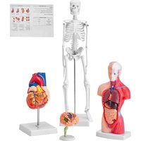
LD-3JT - Vevor - Free user manual and instructions
Find the device manual for free LD-3JT Vevor in PDF.


User questions about LD-3JT Vevor
0 question about this device. Answer the ones you know or ask your own.
Ask a new question about this device
Download the instructions for your in PDF format for free! Find your manual LD-3JT - Vevor and take your electronic device back in hand. On this page are published all the documents necessary for the use of your device. LD-3JT by Vevor.
USER MANUAL LD-3JT Vevor
Technical Support and E-Warranty Certificate www.vevor.com/support
HUMAN BODY TEACHING MODELS USER MANUA
MODEL:LD-3JT
We continue to be committed to provide you tools with competitive price. "Save Half", "Half Price" or any other similar expressions used by us only represents an estimate of savings you might benefit from buying certain tools with us compared to the major top brands and does not necessarily mean to cover all categories of tools offered by us. You are kindly reminded to verify carefully when you are placing an order with us if you are actually saving half in comparison with the top major brands
VEVOR®
TOUGH TOOLS, HALF PRICE
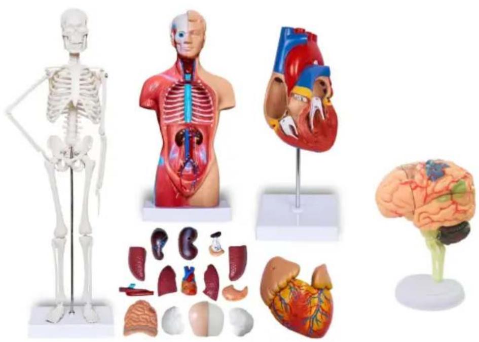
HUMAN BODY TEACHING
MODELS
MODEL:LD-3JT
natural_image
Medical illustrations of human anatomy including skeletal, abdominal, heart, and cerebral structures (no text or labels visible)NEED HELP? CONTACT US!
Have product questions? Need technical support? Please feel from contact us:
Technical Support and E-Warranty Certificate www.vevor.com/support
This is the original instruction, please read all manual instructions carefully before operating. VEVOR reserves a clear interpretation of user manual. The appearance of the product shall be subject to product you received. Please forgive us that we won't inform your use, there are any technology or software updates on our product.
SAFETY INSTRUCTIONS
WARNING:
- Read this instructions before using this product. Failure to do so (result in serious injury.
- After use, please assemble all the parts and keep them vertically.
- Avoid high temperature and direct sunlight.
- Remove minor stains with neutral detergent. The use of alcohol, benzene and other substances may damage the product.
SAVE THESE INSTRUCTIONS
PRODUCT PARAMETER
| Model | LD-3JT | |||
 |  | 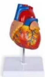 | 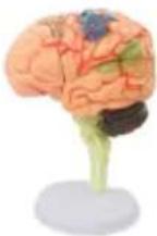 | |
| Labeling Mode | Color Labeling | / | Digital | / |
| Material | PVC | PVC | PVC | PVC |
| Removable Parts | 6 | 15 | 2 | 32 |
INSTRUCTIONS

natural_image
Simple line drawing of a human figure in profile view, no text or symbols presentSkeletal Anatomy
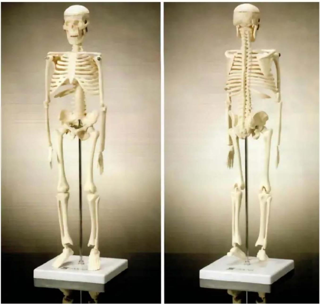
The skeleton has an amazing structure that provides both protection for our vital organs and allows for movement. Check out the figures below learn the identifications of many of the major bones in our bodies.
natural_image
Two identical white skeletal skeleton models displayed on pedestals, showing full skeletal structure and limbs (no text or labels visible)
text_image
Acromion Process Clavicle Ribs 1-12 Ribs 11,12 are floating ribsThere are 12 ribs in the human body, which connect to the sternum front and the vertebral column in the back (see Vertebral Column Fig Below). Ribs 11 and 12 are called floating ribs because they do not an attachment to the sternum in the front. Note the location of the c which is also called the process of the scapula at the other end. co The clavicle attaches to the sternum at one end and to the acromior
text_image
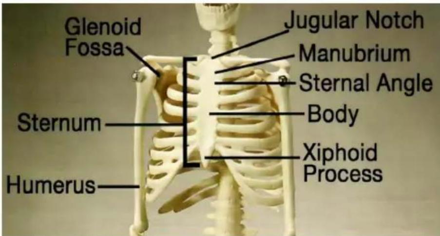
Glenoid Fossa Jugular Notch Manubrium Sternal Angle Body Sternum Xiphoid Process HumerusThe sternum or breast bone is divided into three pieces from top to called the manubrium, the body, and the xiphoid process. The jugular is a small indentation at the top of the sternum, and the sternal ang small groove between the manubrium and the body. The humerus is bone of the upper arm, and it forms the shoulder joint by attaching scapula at the glenoid fossa.
text_image
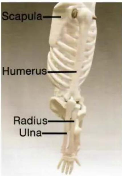
Scapula Humerus Radius UlnaThis figure shows a side view of the upper body so that we can see bones of the arm. The humerus is the bone of the upper arm, and attaches to the scapula to form the shoulder joint. The radius and ul the bones of the forearm, and they attach to the humerus to form the joint. They also attach to the bones of the hand to form the wrist jo
text_image
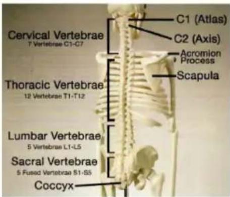
Cervical Vertebrae 7 Vertebrae C1-C7 Thoracic Vertebrae 12 Vertebrae T1-T12 Lumbar Vertebrae 5 Vertebrae L1-L5 Sacral Vertebrae 5 Fused Vertebrae S1-S5 Coccyx C1 (Atlas) C2 (Axis) Acromion Process ScapulaThe vertebral column includes the major bones in the back that prote spinal cord. There are four main divisions of the vertebral column inc 7 cervical vertebrae (numbered C1 to C7), 12 thoracic vertebrae (numbered T1 to T12), 5 lumbar vertebrae (numbered L1 to L5),5 sac vertebrae (numbered S1 to S5), and lastly the coccyx on tailbone. The cervical vertebrae, C1, is called the Atlas,while C2 is known as the A The 5 sacral vertebrae (S1 to S5) are often fused together to form known as the sacrum. Also note the location of the scapula in our b
which is also known as the shoulder blade.
text_image
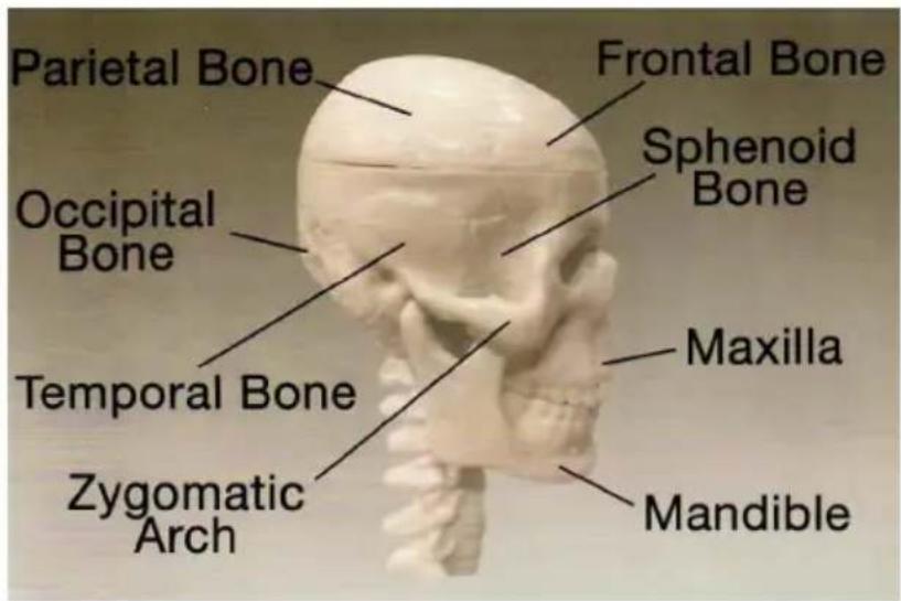
Parietal Bone Frontal Bone Sphenoid Bone Occipital Bone Temporal Bone Maxilla Zygomatic Arch MandibleThe human skull is actually formed from several different bones, many which are attached together through small groves known as sutures (s next Figure). The mandible is the lower jaw bone the maxilla is the jaw bone, and the zygomatic arch represents our cheek bones. The parietal bones, frontal bone, sphenoid bones, temporal bones, and occipital bone make up the top, front, sides, and back of the skull a critical for protecting the brain!
text_image
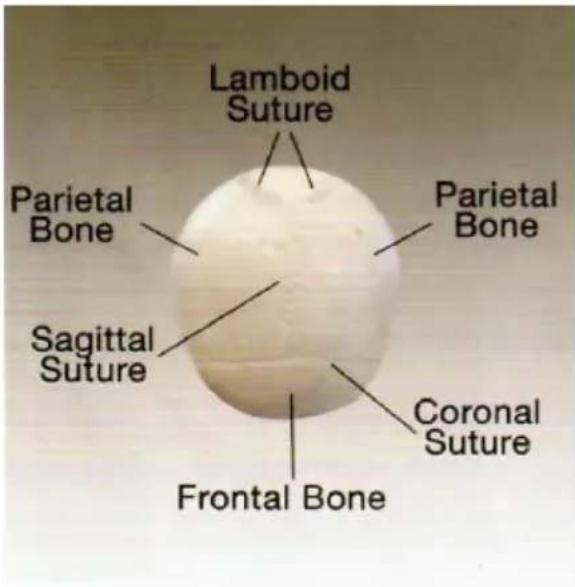
Lamboid Suture Parietal Bone Parietal Bone Sagittal Suture Coronal Suture Frontal BoneWith the exception of the mandible, the bones in the skull are joined another through sutures, which appear as small grooves between these bones. These sutures are actually considered to be joints. However,
most joints in the body, sutures are immovable. Note the location of of the major sutures in the skull above.
text_image
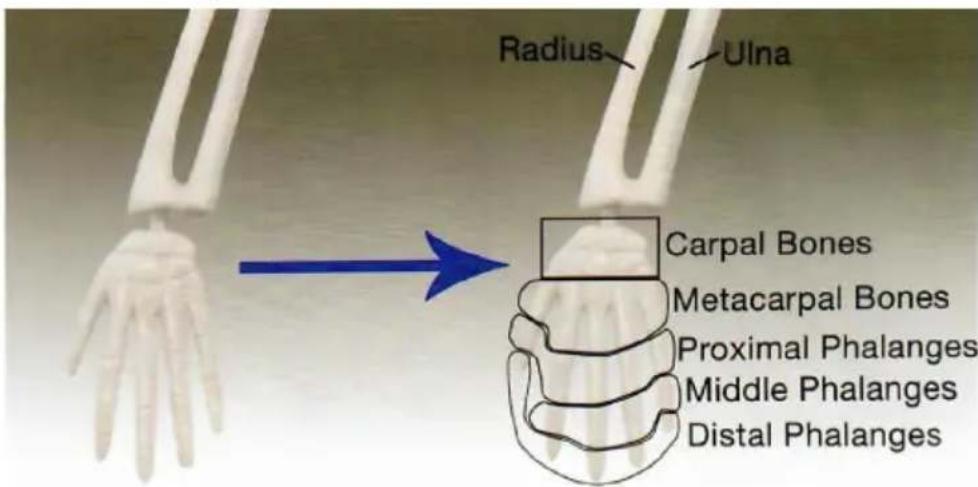
Radius Ulna Carpal Bones Metacarpal Bones Proximal Phalanges Middle Phalanges Distal PhalangesThe hands are formed from multiple small bones. The first bones of hands which are closest to the wrist are called the carpal bones. Ne come the metacarpal bones, and lastly the phalanges. The thumb only two phalanges called the proximal and distal phalanges. However, each the other fingers has three phalanges, named the proximal, middle, and distal phalanges! Note the location of the radius and ulna, which are two bones that form our forearm. The radius is on the same side of as the thumb, which is an easy way to remember its location.
text_image
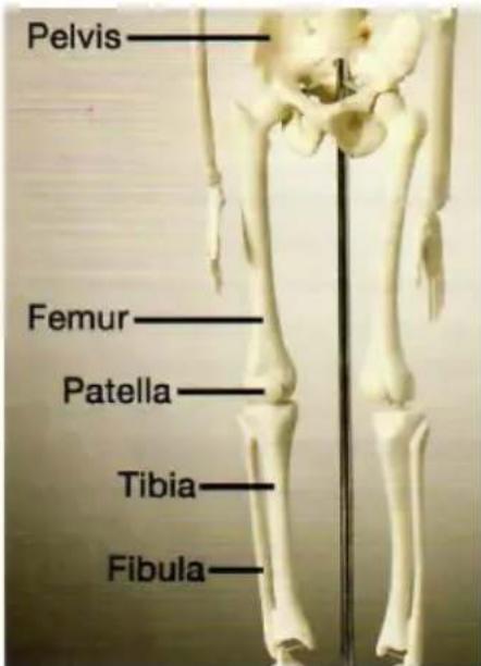
Pelvis Femur Patella Tibia FibulaThe femur is the largest bone in the body and is the bone of our The knee cap is called the patella and our lower legs are made from bones known as the tibia (also known as the shinbone) and the fibu
femur attaches to the pelvis at the hip joint.
text_image
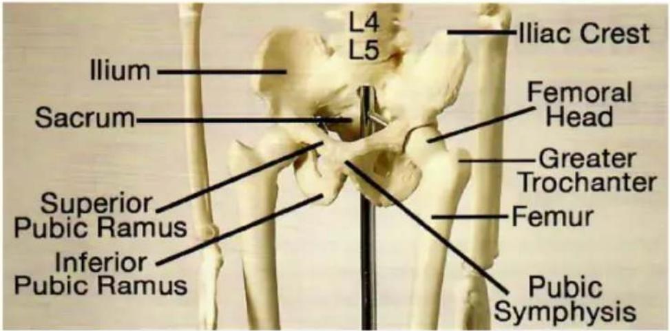
L4 L5 Iliac Crest Ilium Sacrum Superior Pubic Ramus Inferior Pubic Ramus Femoral Head Greater Trochanter Femur Pubic SymphysisThe pelvis is divided into different regions with distinct names. Check some of these regions above including the ilium, iliac crest, superior inferior ramus, and the pubic symphysis. Note that the femur attaches the pelvis to form the hip bone. The top of the femur is called the head and the upper side of the femur is called the greater trochante
text_image
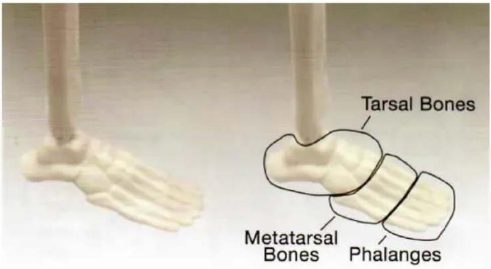
Tarsal Bones Metatarsal Bones PhalangesLike the hands, the feet are also made up of multiple bones. The b that are closest to the ankle joint are known as the tarsal bones. No the metatarsals, followed by the phalanges. Like the thumb, the big t only has two phalanges called the proximal and distal phalanges. How the other toes have three phalanges called the proximal, middle, and phalanges.
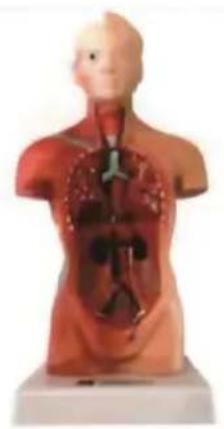
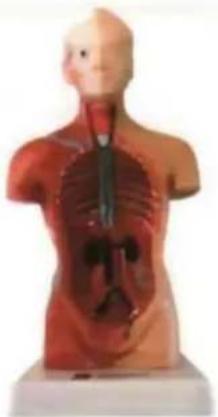
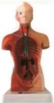
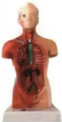
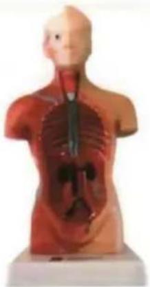
- Lay the Torso Anatomy Model Flat on its Back on a Flat Surface

natural_image
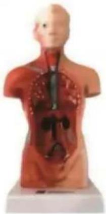
Anatomical model of a human torso showing internal organs including lungs, heart, and intestines (no text or labels visible)- Add the Trachea / Esophagus / Aorta
Piece Between the Lungs
natural_image
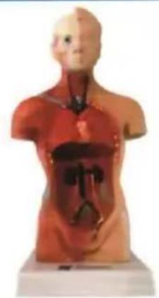
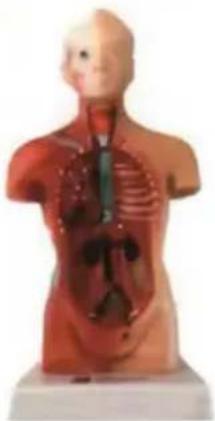
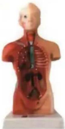
Anatomical model of a human torso showing internal organs and structures (no text or labels visible)- Add the Anterior (Front) Left Lung to the Left Chest
natural_image
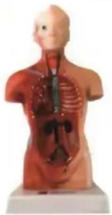
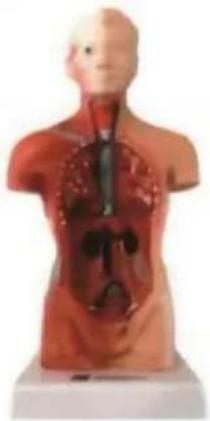
Anatomical model of a human torso showing internal organs (no text or labels visible)- Add the Posterior(Back)Half of the Right Lung to the Right Chest
natural_image
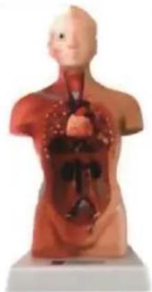
Anatomical model of a human torso showing internal organs and structures (no text or labels visible)- Next Add The Heart on Top of the Trachea Piece
natural_image
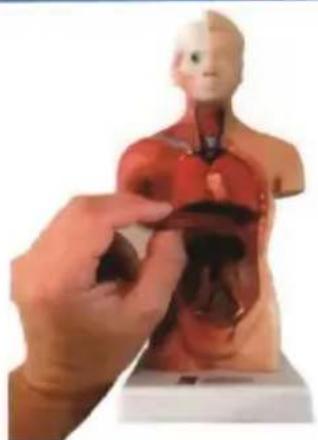
Anatomical model of human body showing internal organs and structures (no text or labels visible)- Add the Diaphragm Piece Right Underneath the Lungs
natural_image
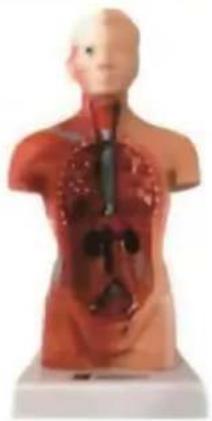
Anatomical model of a human body with red and orange coloring, being held by a hand (no text or labels visible)- Add the Posterior(Back)Half of the Left Lung to the Left Chest
natural_image
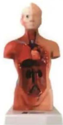
Anatomical model of a human torso showing internal organs and structures (no text or labels visible)- Add the Anterior(Front) Right Lung to the Right Cheae
natural_image
Anatomical model of a human torso showing internal organs (no text or labels visible)- Add the Liver Right Underneath the Diaphragm
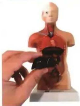
natural_image
Hand holding a small black object next to a red anatomical model of a human torso (no text or symbols visible)- Add the Pancreas / Duodenum / Spleen to Upper Right Abdomen
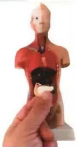
natural_image
Illustration of a human figure in profile view with a hand holding a small object (no text or symbols visible)- CONT. Location of Pancreas / Duodenum/Spleen in Abdomen
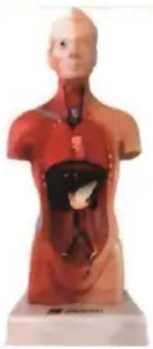
natural_image
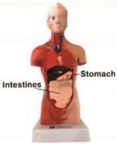
Anatomical model of a human torso and arm, showing internal organs (no text or labels visible)- Add the Intestines and Stomach
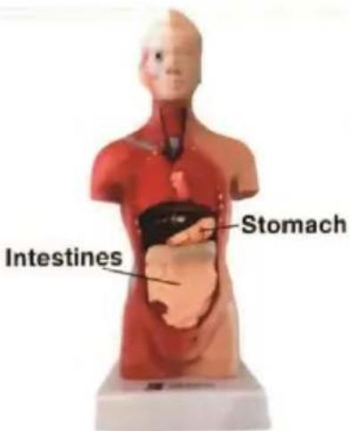
text_image
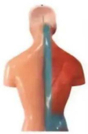
Intestines Stomach- Face the Torso Model Downward on a Flat Surface
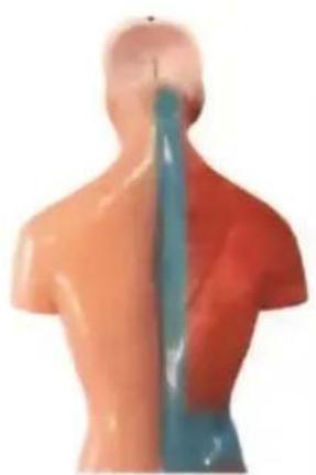
natural_image
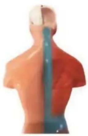
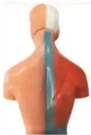
Human back showing a vertical spine with color gradient from orange to blue (no text or symbols)- Add One Half(Hemisphere) of the Brain
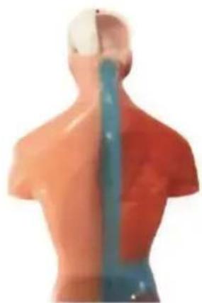
natural_image
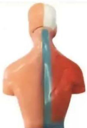
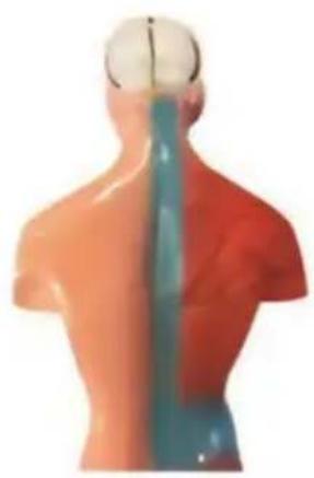
Human back view showing a vertical spine and shoulder (no text or symbols)- Add the Other Half(Hemi-sphere) of the Brain

natural_image
Human back view showing a vertical spine with color gradient from orange to blue (no text or symbols)- Add the Back of the Head
natural_image
Anatomical illustration of a human back showing the spine and shoulder (no text or labels)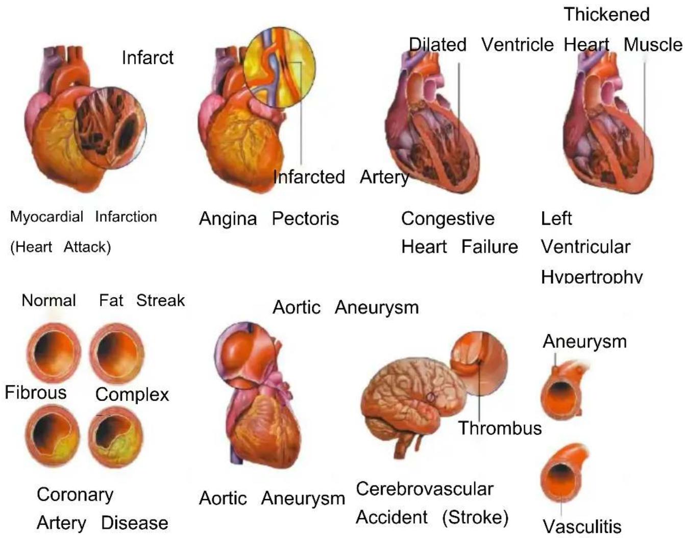
| 1.Right atrium2.Right auricle3.Superior vena cava4.inferior vena cava5.Right ventricle6.Pulmonary trunk | 7.Ascending aorta8.Arch of the aorta9.Brachiocephalic trunk10.Common carotid artery11.Subclavian artery12.Arterial ligament | 13.Descending aorta14.Left auricle15.Left ventricle16.Right coronary artery17.Anterior interventricular branch o left coronary artery |
| 18.Circumflex branch of left coronary artery19.Great cardiac vein20.Middle cardiac vein21.Small cardiac vein22.Posterior interventricular branch or right coronary artery | 23.Coronary sinus24.Fossa ovalis25.Opening of coronary sinus26.Tricuspid valve27.Pulmonary valve28.Left atrium | 29.Bicuspid valve(mitral valve)30.Aortic valve31.Left pulmonary artery32.Left pulmonary vein33.Right pulmonary artery34.Right pulmonary vein |
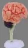
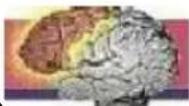
1.Frontal lobe
It is the front area of each cerebral hemisphere and responds to pla problem-solving, emotions, also parts of speech and movement.
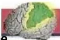
2.Parietal lobe
It is located behind the central sulcus, responsible for perception of to pressure , temperature and pain .
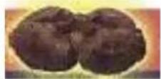
3.Cerebellum
It is located at the back and near the brainstem. It is mainly responsible for motor movements, but also partially for learning, emotions, speaking, and attention

4. Corpus striatum
The group of structures which is also called the “striped body” located in
the depths of each cerebral hemisphere
natural_image
Abstract illustration of a stylized figure with a flowing tail, set against a red background (no text or symbols)5.Ventricle
These are hollow chambers filled with cerebrospinal fluid supporting the upper tissues of the brain
natural_image
Close-up of a textured, irregularly shaped food item with a visible interior area (no text or symbols)6.Occipital lobe
It is the rear part of cerebrum responsible for vision

7. Corpus Callosum
It is a thick band of millions of nerve fibers located deeply in the cthe brain
8. Insula
A pair of left and right insula which is located deeply in the brain, as important for the recognition of disgust
9.Brainstem
It is a lower extension of the brain and connects to the spinal cord. mainly responsible for breathing , digestion , heart rate and blood pre control
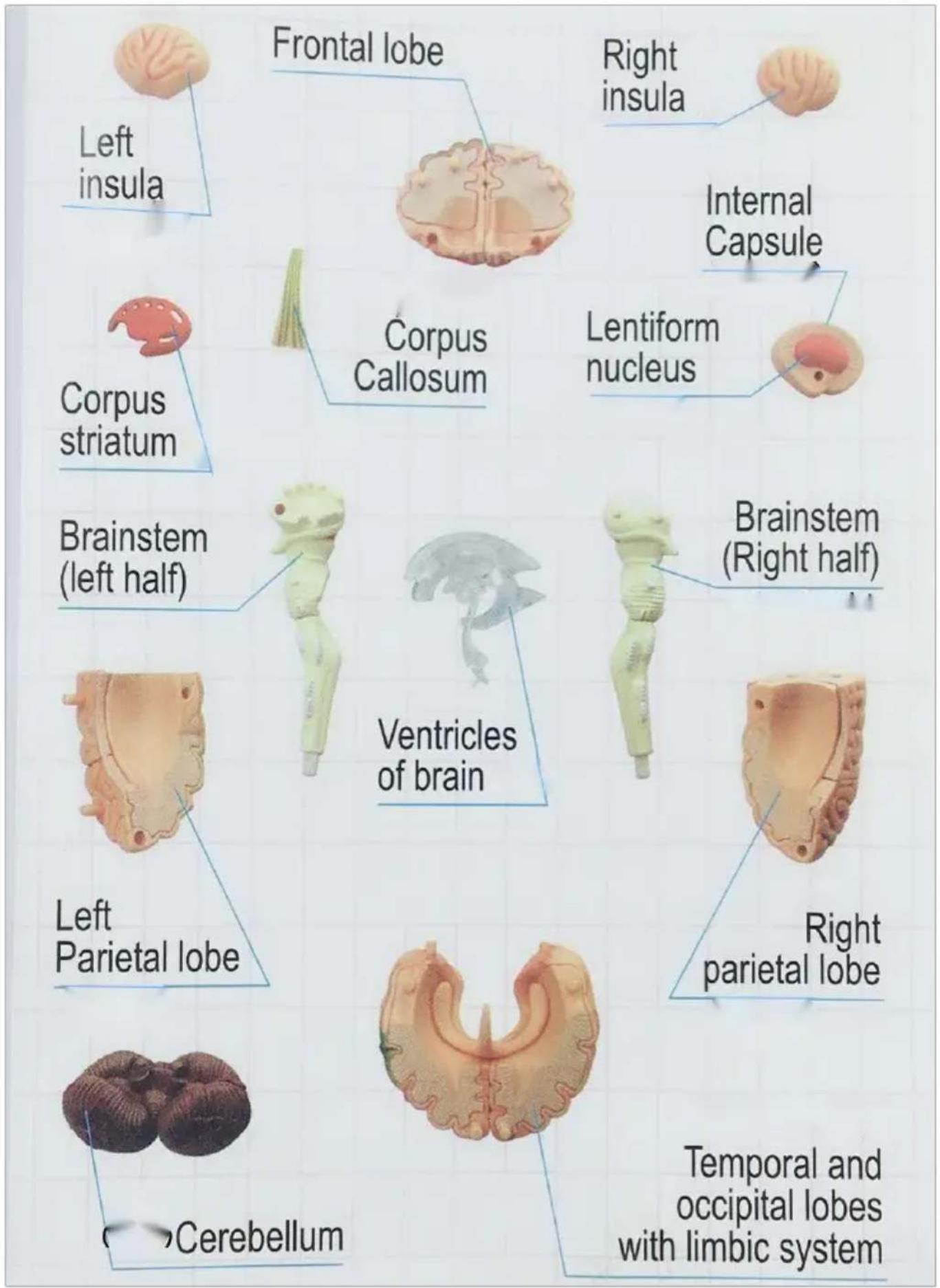
text_image
Left insula Frontal lobe Right insula Corpus striatum Corpus Callosum Internal Capsule Lentiform nucleus Brainstem (left half) Brainstem (Right half) Ventricles of brain Left Parietal lobe Right parietal lobe Temporal and occipital lobes with limbic system Cerebellum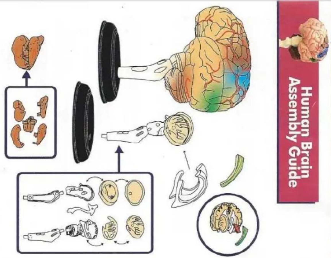
flowchart
graph TD
    A["Feeding"] --> B["Carbohydrates"]
    B --> C["Carbohydrates in brain"]
    C --> D["Carbohydrates in ears"]
    D --> E["Carbohydrates in ears in brain"]
    E --> F["Carbohydrates in ears in ears"]
    F --> G["Carbohydrates in ears in ears"]
    G --> H["Carbohydrates in ears in ears"]
    H --> I["Carbohydrates in ears in ears"]
    I --> J["Carbohydrates in ears in ears"]
    J --> K["Carbohydrates in ears in ears"]
    K --> L["Carbohydrates in ears in ears"]
    L --> M["Carbohydrates in ears in ears"]
    M --> N["Carbohydrates in ears in ears"]
    N --> O["Carbohydrates in ears in ears"]
    O --> P["Carbohydrates in ears in ears"]
    P --> Q["Carbohydrates in ears in ears"]
    Q --> R["Carbohydrates in ears in ears"]
    R --> S["Carbohydrates in ears in ears"]
natural_image
Close-up of a decorative floral or coral-like object with a dark base and small white elements against a solid red background (no text or symbols visible)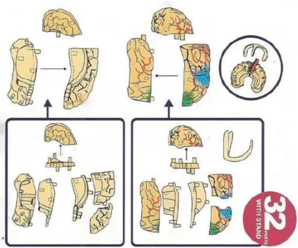
flowchart
graph TD
    A["Brain anatomy"] --> B["Anatomical change"]
    B --> C["Anatomical change with color-coded regions"]
    C --> D["Anatomical change with circled area"]
    D --> E["Anatomical change with L-shaped region"]
    E --> F["Anatomical change with cross-sections"]
    F --> G["Anatomical change with left side of L-shaped region"]
    G --> H["Anatomical change with right side of L-shaped region"]
    style A fill:#f9f,stroke:#333
    style B fill:#f9f,stroke:#333
    style C fill:#f9f,stroke:#333
    style D fill:#f9f,stroke:#333
    style E fill:#f9f,stroke:#333
    style F fill:#f9f,stroke:#333
    style G fill:#f9f,stroke:#333
    style H fill:#f9f,stroke:#333
Manufacturer: Shanghaimuxinmuyeyouxiangongsi
Address: Shuangchenglu 803nong11hao1602A-1609shi, baoshanqu, shanghai 200000 CN.
Imported to AUS: SIHAO PTY LTD. 1 ROKEVA STREET-EASTWOOD NSW 2122 Australia
Imported to USA: Sanven Technology Ltd. Suite 250, 9166 Anaheim Place, Rancho Cucamonga, CA 91730
| EC | REP |
YH CONSULTING LIMITED. C/O YH Consulting Limited Office 147, Centurion House, London Road, Staines-upon-Thames, Surrey, TW18 4AX
VEVOR®
TOUGH TOOLS, HALF PRICE
Technical Support and E-Warranty Certificate
www.vevor.com/support
VEVOR®
TOUGH TOOLS, HALF PRICE
natural_image
Medical illustrations of human anatomy including skeletal, skeletal muscle, internal organs, and cerebral blood vessels (no text or labels visible)BESOIN D'AIDE? CONTACTEZ-NOUS!
natural_image
Simple line drawing of a human figure standing in front of a gray rectangular background (no text or symbols)Anatomie du
natural_image
Two identical white skeletal models displayed on pedestals, showing full skeletal structure and limbs (no text or labels visible)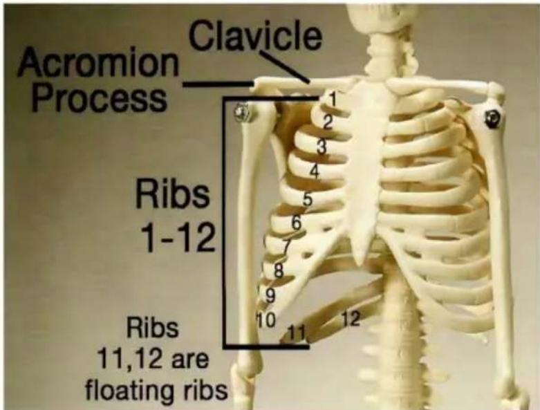
text_image
Acromion Process Clavicle Ribs 1-12 Ribs 11,12 are floating ribstext_image
Parietal Bone Frontal Bone Sphenoid Bone Occipital Bone Temporal Bone Maxilla Zygomatic Arch Mandibletext_image
Lamboid Suture Parietal Bone Parietal Bone Sagittal Suture Coronal Suture Frontal Bone- Lay the Torso Anatomy Model Flat on its Back on a Flat Surface
natural_image
Anatomical model of a human torso showing internal organs and structures (no text or labels visible)- Add the Posterior(Back)Half of the Right Lung to the Right Chest
natural_image
Anatomical model of a human torso showing internal organs and structures (no text or labels visible)- Add the Posterior(Back)Half of the Left Lung to the Left Chest
natural_image
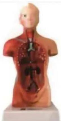
Anatomical model of a human torso showing internal organs (no text or labels visible)- Add the Trachea / Esophagus / Aorta
Piece Between the Lungs
natural_image
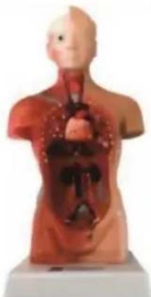
Anatomical model of a human torso and arm, showing internal organs (no text or labels visible)- Next Add The Heart on Top of the Trachea Piece
natural_image
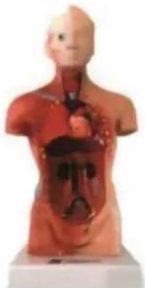
Anatomical model of a human torso showing internal organs (no text or labels visible)- Add the Anterior(Front) Right Lung to the Right Cheae
natural_image
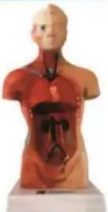
Anatomical model of a human torso and arm, showing internal organs (no text or labels visible)- Add the Anterior (Front) Left Lung to the Left Chest
natural_image
Anatomical model of a human torso showing internal organs (no text or labels visible)- Add the Diaphragm Piece Right Underneath the Lungs
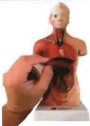
natural_image
Hand holding a model of a human body in profile view (no text or symbols visible)- Add the Liver Right Underneath the Diaphragm
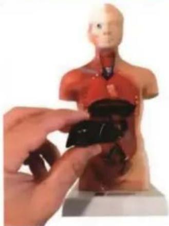
natural_image
Hand holding a small black object next to a transparent model of a human torso (no text or symbols visible)- Add the Pancreas / Duodenum / Spleen to Upper Right Abdomen
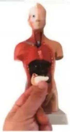
natural_image
Illustration of a human figure with a hand holding an object, no visible text or symbols- CONT. Location of Pancreas / Duodenum/Spleen in Abdomen
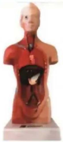
natural_image
Anatomical model of a human torso and arm, showing internal organs (no text or labels visible)- Add the Intestines and Stomach
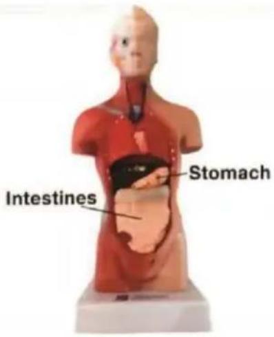
text_image
Intestines Stomach- Face the Torso Model Downward on a Flat Surface
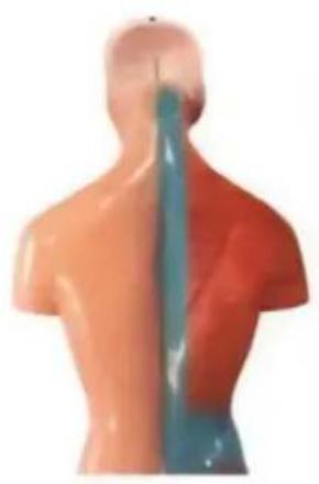
natural_image
Human back view showing a vertical spine with colored muscle fibers (no text or labels)- Add One Half(Hemisphere) of the Brain
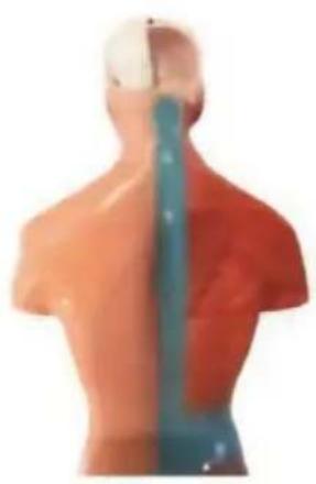
natural_image
Human back view showing a vertical spine with colored regions (no text or symbols)- Add the Other Half(Hemi-sphere) of the Brain
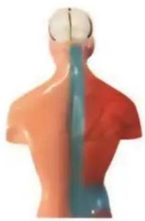
natural_image
Back view of a human torso with a vertical blue surgical band on the shoulder area (no text or symbols)- Add the Back of the Head
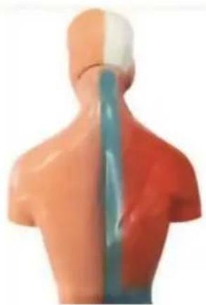
natural_image
Anatomical illustration of a human back and shoulder showing muscle fibers (no text or labels)
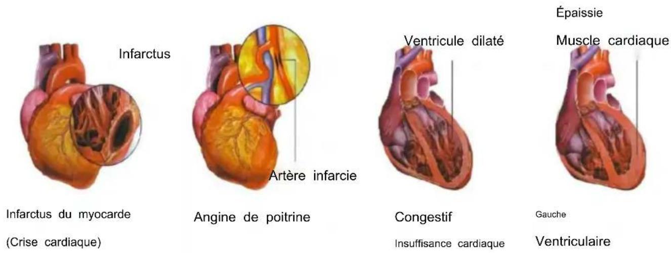
Hypertrophie
Plaque

Plaque
Coronaire
Maladie artérielle
natural_image
Anatomical illustration of the human heart and major blood vessels (no text or labels)natural_image
Abstract illustration of a stylized figure with flowing hair against a red background (no text or symbols)5. Ventricule
natural_image
Close-up of a textured, irregularly shaped object with a visible interior area (no text or symbols)text_image
Left insula Frontal lobe Right insula Corpus striatum Corpus Callosum Internal Capsule Lentiform nucleus Brainstem (left half) Ventricles of brain Brainstem (Right half) Left Parietal lobe Right parietal lobe Cerebellum Temporal and occipital lobes with limbic system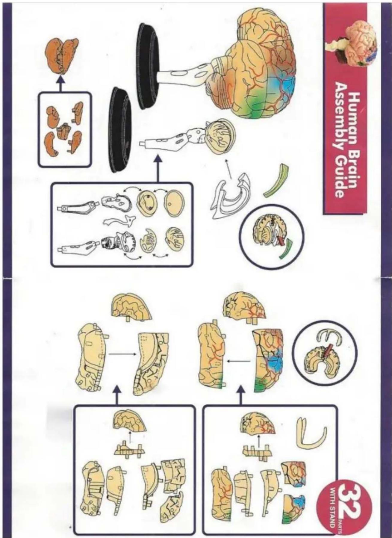
flowchart
graph TD
    A["Feeding"] --> B["Assembly"]
    B --> C["Brain with Human Brain"]
    C --> D["Assembly with Human Brain"]
    D --> E["Assembly with Human Brain"]
    E --> F["Assembly with Human Brain"]
    F --> G["Assembly with Human Brain"]
    G --> H["Assembly with Human Brain"]
    H --> I["Assembly with Human Brain"]
    I --> J["Assembly with Human Brain"]
    J --> K["Assembly with Human Brain"]
    K --> L["Assembly with Human Brain"]
    L --> M["Assembly with Human Brain"]
    M --> N["Assembly with Human Brain"]
    N --> O["Assembly with Human Brain"]
    O --> P["Assembly with Human Brain"]
    P --> Q["Assembly with Human Brain"]
    Q --> R["Assembly with Human Brain"]
    R --> S["Assembly with Human Brain"]
    S --> T["Assembly with Human Brain"]
    T --> U["Assembly with Human Brain"]
    U --> V["Assembly with Human Brain"]
    V --> W["Assembly with Human Brain"]
    W --> X["Assembly with Human Brain"]
    X --> Y["Assembly with Human Brain"]
    Y --> Z["Assembly with Human Brain"]
    Z --> AA["Assembly with Human Brain"]
    AA --> AB["Assembly with Human Brain"]
    AB --> AC["Assembly with Human Brain"]
    AC --> AD["Assembly with Human Brain"]
    AD --> AE["Assembly with Human Brain"]
    AE --> AF["Assembly with Human Brain"]
    AF --> AG["Assembly with Human Brain"]
    AG --> AH["Assembly with Human Brain"]
    AH --> AI["Assembly with Human Brain"]
    AI --> AJ["Assembly with Human Brain"]
    AJ --> AK["Assembly with Human Brain"]
    AK --> AL["Assembly with Human Brain"]
    AL --> AM["Assembly with Human Brain"]
    AM --> AN["Assembly with Human Brain"]
    AN --> AO["Assembly with Human Brain"]
    AO --> AP["Assembly with Human Brain"]
    AP --> AQ["Assembly with Human Brain"]
    AQ --> AR["Assembly with Human Brain"]
    AR --> AS["Assembly with Human Brain"]
    AS --> AT["Assembly with Human Brain"]
    AT --> AU["Assembly with Human Brain"]
    AU --> AV["Assembly with Human Brain"]
    AV --> AW["Assembly with Human Brain"]
    AW --> AX["Assembly with Human Brain"]
    AX --> AY["Assembly with Human Brain"]
Manufacturer: Shanghaimuxinmuyeyouxiangongsi Address: Shuangchenglu 803nong11hao1602A-1609shi, baoshanqu, shanghai 200000 CN. Imported to AUS: SIHAO PTY LTD. 1 ROKEVA STREET- EASTWOOD NSW 2122 Australia Imported to USA: Sanven Technology Ltd. Suite 250, 9166 Anaheim Place, Rancho Cucamonga, CA 91730

YH CONSULTING LIMITED. C/O YH Consulting Limited Office 147, Centurion House, London Road, Staines-upon-Thames, Surrey, TW18 4AX
VEVOR®
TOUGH TOOLS, HALF PRICE
natural_image
Medical illustrations of human anatomy including skeletal, skeletal muscle, internal organs, and cerebral blood vessels (no text or labels visible)natural_image
Simple line drawing of a human skeleton in profile view, no text or symbols presentAnatomie des Skeletts
natural_image
Two identical white skeletal skeleton models displayed on pedestals, showing full skeletal structure and limbs (no text or labels visible)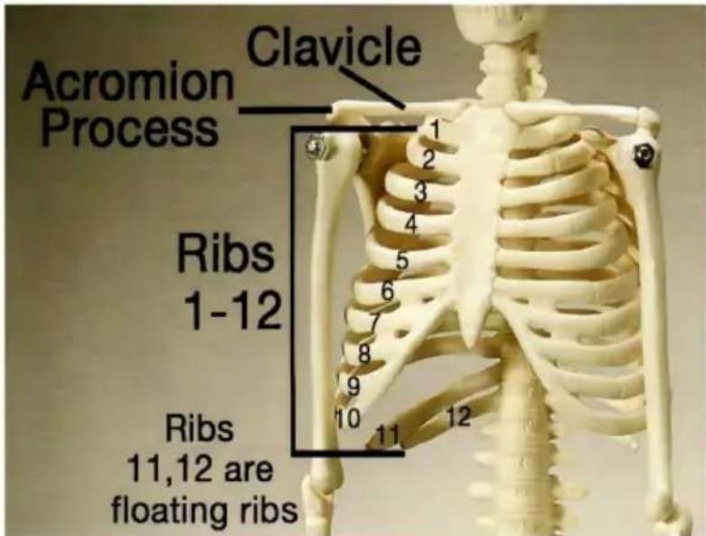
text_image
Acromion Process Clavicle Ribs 1-12 Ribs 11,12 are floating ribstext_image
Parietal Bone Frontal Bone Sphenoid Bone Occipital Bone Temporal Bone Maxilla Zygomatic Arch Mandibletext_image
Lamboid Suture Parietal Bone Parietal Bone Sagittal Suture Coronal Suture Frontal Bone- Lay the Torso Anatomy Model Flat on its Back on a Flat Surface
natural_image
Anatomical model of a human torso showing internal organs and structures (no text or labels visible)- Add the Posterior(Back)Half of the Right Lung to the Right Chest
natural_image
Anatomical model of a human torso showing internal organs and structures (no text or labels visible)- Add the Posterior(Back)Half of the Left Lung to the Left Chest
natural_image
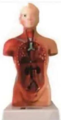
Anatomical model of a human torso showing internal organs (no text or labels visible)- Add the Trachea / Esophagus / Aorta
Piece Between the Lungs
natural_image
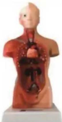
Anatomical model of a human torso and arm, showing internal organs (no text or labels visible)- Next Add The Heart on Top of the Trachea Piece
natural_image
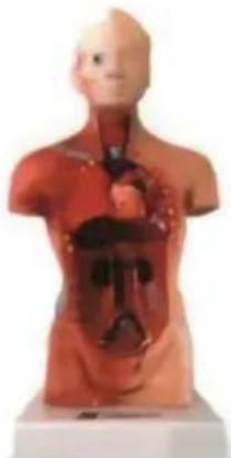
Anatomical model of a human torso showing internal organs (no text or labels visible)- Add the Anterior(Front) Right Lung to the Right Cheae
natural_image
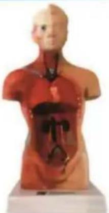
Anatomical model of a human torso and arm, showing internal organs (no text or labels visible)- Add the Anterior (Front) Left Lung to the Left Chest
natural_image
Anatomical model of a human torso showing internal organs (no text or labels visible)- Add the Diaphragm Piece Right Underneath the Lungs
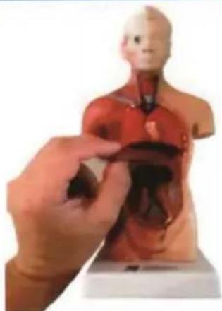
natural_image
Hand holding a model of a human body in profile view (no text or symbols visible)- Add the Liver Right Underneath the Diaphragm
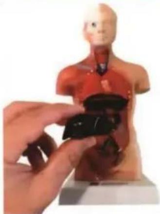
natural_image
Hand holding a small black object next to a transparent model of a human torso (no text or symbols visible)- Add the Pancreas / Duodenum / Spleen to Upper Right Abdomen
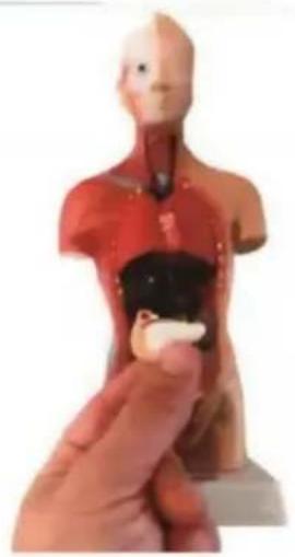
natural_image
Illustration of a human figure with a hand holding an object, no visible text or symbols- CONT. Location of Pancreas / Duodenum/Spleen in Abdomen
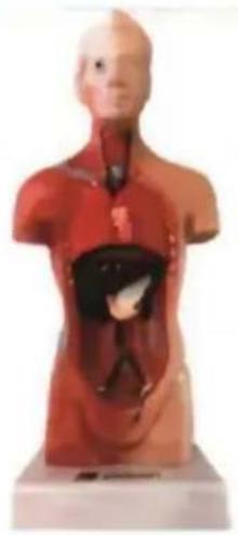
natural_image
Anatomical model of a human torso and arm, showing internal organs (no text or labels visible)- Add the Intestines and Stomach
text_image
Intestines Stomach- Face the Torso Model Downward on a Flat Surface
natural_image
Human back view showing a vertical spine with colored muscle fibers (no text or labels)- Add One Half(Hemisphere) of the Brain
natural_image
Human back view showing a vertical spine with colored regions (no text or symbols)- Add the Other Half(Hemi-sphere) of the Brain
natural_image
Back view of a human torso with a vertical blue surgical band on the shoulder area (no text or symbols)- Add the Back of the Head
natural_image
Anatomical model of a human back showing internal muscle fibers (no text or labels)
natural_image
Abstract illustration of a stylized figure with flowing hair against a red background (no text or symbols)5.Ventrikel
natural_image
Close-up of a textured, irregularly shaped object with a visible interior area (no text or symbols)6.Occipitallappen Es
text_image
Left insula Frontal lobe Right insula Corpus striatum Corpus Callosum Internal Capsule Lentiform nucleus Brainstem (left half) Ventricles of brain Brainstem (Right half) Left Parietal lobe Right parietal lobe Cerebellum Temporal and occipital lobes with limbic system
flowchart
graph TD
    A["Feeding"] --> B["Assembly"]
    B --> C["Brain with Human Brain"]
    C --> D["Assembly with Human Brain"]
    D --> E["Assembly with Human Brain"]
    E --> F["Assembly with Human Brain"]
    F --> G["Assembly with Human Brain"]
    G --> H["Assembly with Human Brain"]
    H --> I["Assembly with Human Brain"]
    I --> J["Assembly with Human Brain"]
    J --> K["Assembly with Human Brain"]
    K --> L["Assembly with Human Brain"]
    L --> M["Assembly with Human Brain"]
    M --> N["Assembly with Human Brain"]
    N --> O["Assembly with Human Brain"]
    O --> P["Assembly with Human Brain"]
    P --> Q["Assembly with Human Brain"]
    Q --> R["Assembly with Human Brain"]
    R --> S["Assembly with Human Brain"]
    S --> T["Assembly with Human Brain"]
    T --> U["Assembly with Human Brain"]
    U --> V["Assembly with Human Brain"]
    V --> W["Assembly with Human Brain"]
    W --> X["Assembly with Human Brain"]
    X --> Y["Assembly with Human Brain"]
    Y --> Z["Assembly with Human Brain"]
    Z --> AA["Assembly with Human Brain"]
    AA --> AB["Assembly with Human Brain"]
    AB --> AC["Assembly with Human Brain"]
    AC --> AD["Assembly with Human Brain"]
    AD --> AE["Assembly with Human Brain"]
    AE --> AF["Assembly with Human Brain"]
    AF --> AG["Assembly with Human Brain"]
    AG --> AH["Assembly with Human Brain"]
    AH --> AI["Assembly with Human Brain"]
    AI --> AJ["Assembly with Human Brain"]
    AJ --> AK["Assembly with Human Brain"]
    AK --> AL["Assembly with Human Brain"]
    AL --> AM["Assembly with Human Brain"]
    AM --> AN["Assembly with Human Brain"]
    AN --> AO["Assembly with Human Brain"]
    AO --> AP["Assembly with Human Brain"]
    AP --> AQ["Assembly with Human Brain"]
    AQ --> AR["Assembly with Human Brain"]
    AR --> AS["Assembly with Human Brain"]
    AS --> AT["Assembly with Human Brain"]
    AT --> AU["Assembly with Human Brain"]
    AU --> AV["Assembly with Human Brain"]
    AV --> AW["Assembly with Human Brain"]
    AW --> AX["Assembly with Human Brain"]
    AX --> AY["Assembly with Human Brain"]
Manufacturer: Shanghaimuxinmuyeyouxiangongsi Address: Shuangchenglu 803nong11hao1602A-1609shi, baoshanqu, shanghai 200000 CN. Imported to AUS: SIHAO PTY LTD. 1 ROKEVA STREET- EASTWOOD NSW 2122 Australia Imported to USA: Sanven Technology Ltd. Suite 250, 9166 Anaheim Place, Rancho Cucamonga, CA 91730

YH CONSULTING LIMITED. C/O YH Consulting Limited Office 147, Centurion House, London Road, Staines-upon-Thames, Surrey, TW18 4AX
VEVOR®
TOUGH TOOLS, HALF PRICE
www.vevor.com/support
VEVOR®
TOUGH TOOLS, HALF PRICE
natural_image
Medical illustrations of human anatomy including skeletal, skeletal muscle, internal organs, and cerebral blood vessels (no text or labels visible)natural_image
Simple line drawing of a human skeleton in profile view, no text or symbols presentAnatomia
natural_image
Two identical white skeletal skeleton models displayed on pedestals, showing full skeletal structure and limbs (no text or labels visible)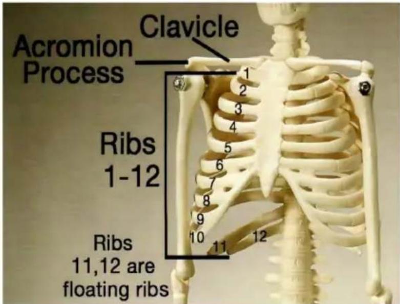
text_image
Acromion Process Clavicle Ribs 1-12 Ribs 11,12 are floating ribstext_image
Parietal Bone Frontal Bone Occipital Bone Sphenoid Bone Temporal Bone Maxilla Zygomatic Arch Mandibletext_image
Lamboid Suture Parietal Bone Parietal Bone Sagittal Suture Coronal Suture Frontal Bone- Lay the Torso Anatomy Model Flat on its Back on a Flat Surface
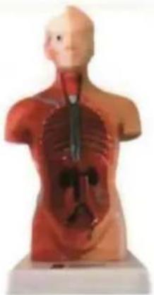
natural_image
Anatomical model of a human torso showing internal organs and structures (no text or labels visible)- Add the Posterior(Back)Half of the Right Lung to the Right Chest
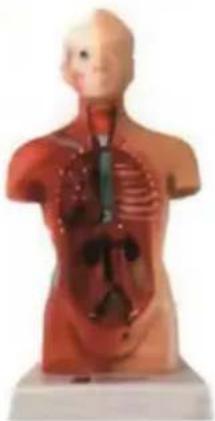
natural_image
Anatomical model of a human torso showing internal organs and structures (no text or labels visible)- Add the Posterior(Back)Half of the Left Lung to the Left Chest
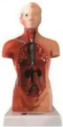
natural_image
Anatomical model of a human torso showing internal organs (no text or labels visible)- Add the Trachea / Esophagus / Aorta
Piece Between the Lungs
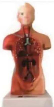
natural_image
Anatomical model of a human torso and arm, showing internal organs (no text or labels visible)- Next Add The Heart on Top of the Trachea Piece

natural_image
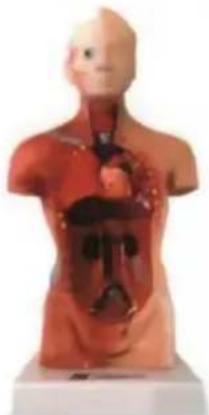
Anatomical model of a human torso showing internal organs (no text or labels visible)- Add the Anterior(Front) Right Lung to the Right Cheae
natural_image
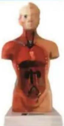
Anatomical model of a human torso and arm, showing internal organs (no text or labels visible)- Add the Anterior (Front) Left Lung to the Left Chest
natural_image
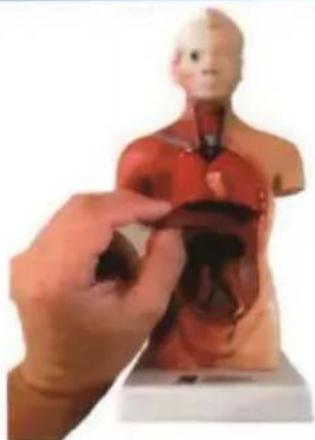
Anatomical model of a human torso showing internal organs (no text or labels visible)- Add the Diaphragm Piece Right Underneath the Lungs
natural_image
Hand holding a model of a human body in profile view (no text or symbols visible)- Add the Liver Right Underneath the Diaphragm

natural_image
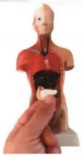
Hand holding a small black object next to a transparent model of a human torso (no text or symbols visible)- Add the Pancreas / Duodenum / Spleen to Upper Right Abdomen
natural_image
Illustration of a human figure with a hand holding an object, no visible text or symbols- CONT. Location of Pancreas / Duodenum/Spleen in Abdomen
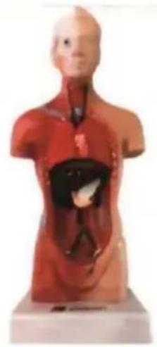
natural_image
Anatomical model of a human torso and arm, showing internal organs (no text or labels visible)- Add the Intestines and Stomach
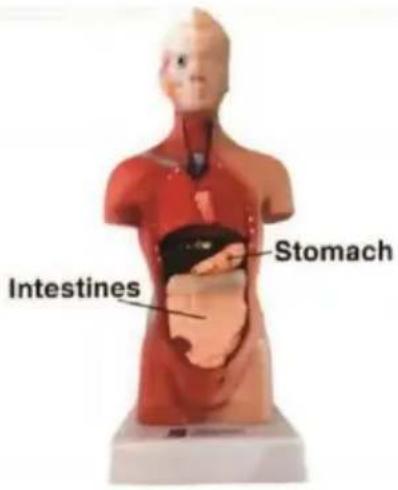
text_image
Intestines Stomach- Face the Torso Model Downward on a Flat Surface
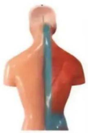
natural_image
Human back view showing a vertical spine with colored muscle fibers (no text or labels)- Add One Half(Hemisphere) of the Brain
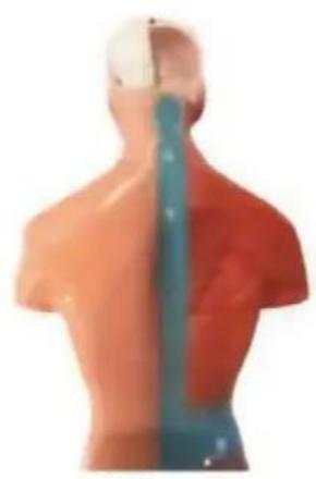
natural_image
Human back view showing a vertical spine with color gradient from orange to blue (no text or symbols)- Add the Other Half(Hemi-sphere) of the Brain
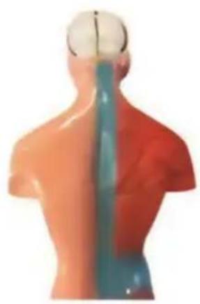
natural_image
Back view of a human torso with a vertical blue surgical band on the shoulder area (no text or symbols)- Add the Back of the Head
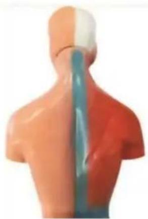
natural_image
Anatomical model of a human back showing internal muscle fibers (no text or labels)
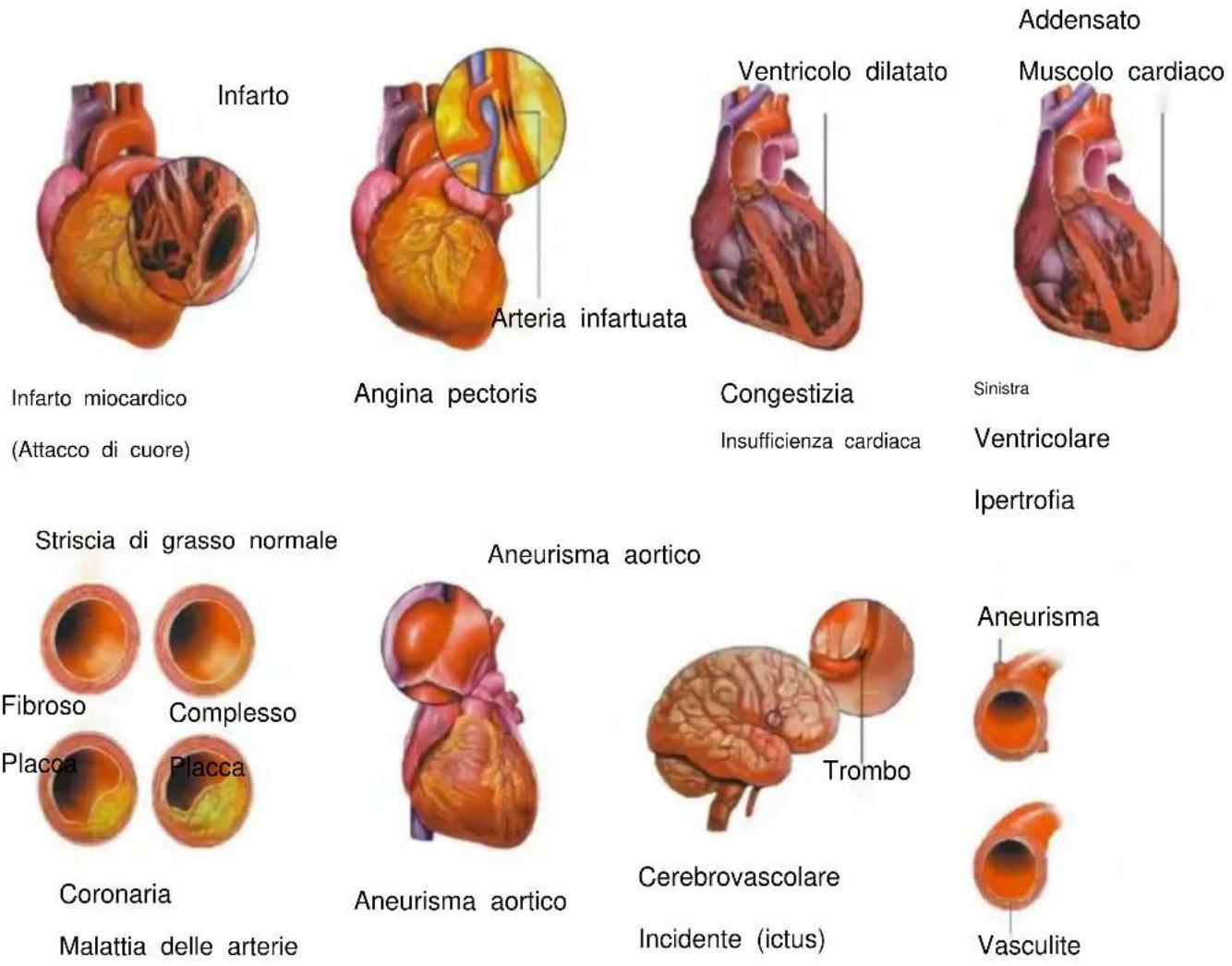
- Atrio destro 2.
Auricola destra 3.
Vena cava superiore
natural_image
Abstract illustration of a stylized figure with flowing hair against a red background (no text or symbols)5.Ventricolo
natural_image
Close-up of a textured, irregularly shaped object with a visible interior area (no text or symbols)6. Lobo occipitale
text_image
Left insula Frontal lobe Right insula Corpus striatum Corpus Callosum Internal Capsule Lentiform nucleus Brainstem (left half) Ventricles of brain Brainstem (Right half) Left Parietal lobe Right parietal lobe Cerebellum Temporal and occipital lobes with limbic system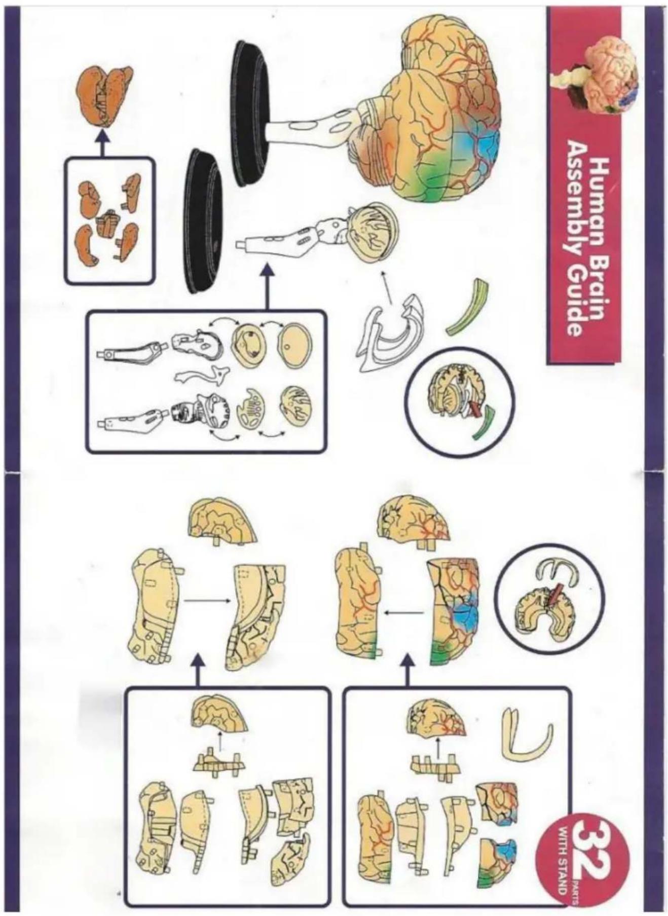
flowchart
graph TD
    A["Feeding"] --> B["Assembly"]
    B --> C["Brain Assembly"]
    C --> D["Human Brain Assembly Guide"]
    D --> E["Assembly"]
    E --> F["Final Assembly"]
    subgraph Human_Brain_1
        G["Red brain icon"] --> H["Human Brain icon"]
        I["Green brain icon"] --> J["Human Brain icon"]
        K["Blue brain icon"] --> L["Human Brain icon"]
        M["White brain icon"] --> N["Human Brain icon"]
        O["Black brain icon"] --> P["Human Brain icon"]
        Q["Yellow brain icon"] --> R["Human Brain icon"]
        S["Green brain icon"] --> T["Human Brain icon"]
        U["Blue brain icon"] --> V["Human Brain icon"]
        W["White brain icon"] --> X["Human Brain icon"]
        Y["Black brain icon"] --> Z["Human Brain icon"]
        AA["Yellow brain icon"] --> AB["Human Brain icon"]
        AC["Green brain icon"] --> AD["Human Brain icon"]
        AE["Blue brain icon"] --> AF["Human Brain icon"]
        AG["White brain icon"] --> AH["Human Brain icon"]
        AI["Black brain icon"] --> AJ["Human Brain icon"]
        AK["Yellow brain icon"] --> AL["Human Brain icon"]
        AM["Green brain icon"] --> AN["Human Brain icon"]
        AO["Blue brain icon"] --> AP["Human Brain icon"]
        AQ["White brain icon"] --> AR["Human Brain icon"]
        AS["Black brain icon"] --> AT["Human Brain icon"]
        AU["Yellow brain icon"] --> AV["Human Brain icon"]
        AW["Green brain icon"] --> AX["Human Brain icon"]
        AY["Red brain icon"] --> AZ["Human Brain Icon"]
    end
Manufacturer: Shanghaimuxinmuyeyouxiangongsi Address: Shuangchenglu 803nong11hao1602A-1609shi, baoshanqu, shanghai 200000 CN. Imported to AUS: SIHAO PTY LTD. 1 ROKEVA STREET- EASTWOOD NSW 2122 Australia Imported to USA: Sanven Technology Ltd. Suite 250, 9166 Anaheim Place, Rancho Cucamonga, CA 91730

YH CONSULTING LIMITED. C/O YH Consulting Limited Office 147, Centurion House, London Road, Staines-upon-Thames, Surrey, TW18 4AX
VEVOR®
TOUGH TOOLS, HALF PRICE
elettronica www.vevor.com/support
VEVOR®
TOUGH TOOLS, HALF PRICE
natural_image
Medical illustrations of human anatomy including skeletal, skeletal muscle, internal organs, and brain models (no text or labels)natural_image
Simple line drawing of a human skeleton in profile view, no text or symbols presentAnatomía del
natural_image
Two identical white skeletal skeleton models displayed on pedestals, showing full skeletal structure and limbs (no text or labels visible)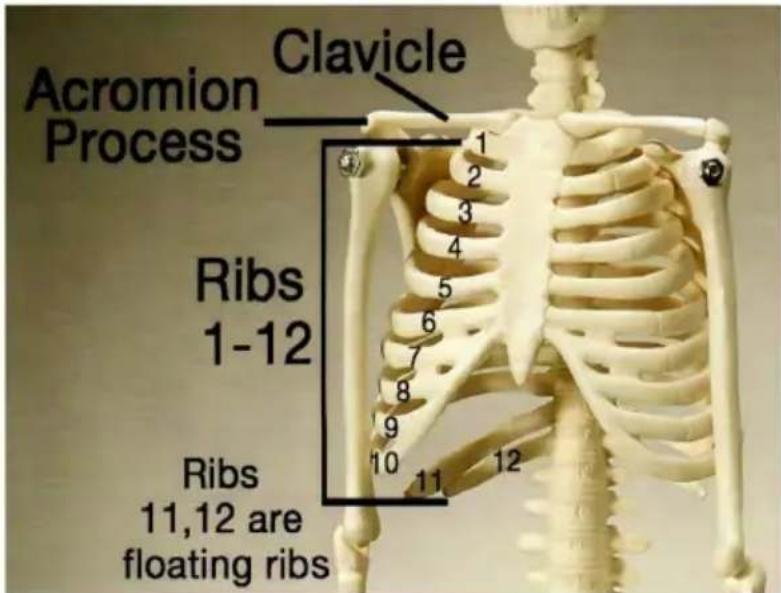
text_image
Acromion Process Clavicle Ribs 1-12 Ribs 11,12 are floating ribstext_image
Parietal Bone Frontal Bone Occipital Bone Sphenoid Bone Temporal Bone Maxilla Zygomatic Arch Mandibletext_image
Lamboid Suture Parietal Bone Parietal Bone Sagittal Suture Coronal Suture Frontal Bone- Lay the Torso Anatomy Model Flat on its Back on a Flat Surface
natural_image
Anatomical model of a human torso showing internal organs and structures (no text or labels visible)- Add the Posterior(Back)Half of the Right Lung to the Right Chest
natural_image
Anatomical model of a human torso showing internal organs and structures (no text or labels visible)- Add the Posterior(Back)Half of the Left Lung to the Left Chest
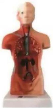
natural_image
Anatomical model of a human torso showing internal organs (no text or labels visible)- Add the Trachea / Esophagus / Aorta
Piece Between the Lungs
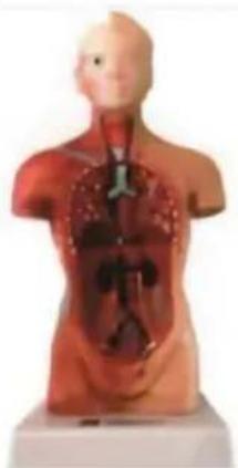
natural_image
Anatomical model of a human torso and arm, showing internal organs (no text or labels visible)- Next Add The Heart on Top of the Trachea Piece
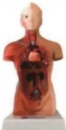
natural_image
Anatomical model of a human torso showing internal organs (no text or labels visible)- Add the Anterior(Front) Right Lung to the Right Cheae
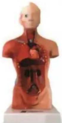
natural_image
Anatomical model of a human torso and arm, showing internal organs (no text or labels visible)- Add the Anterior (Front) Left Lung to the Left Chest
natural_image
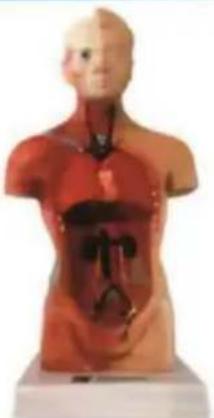
Anatomical model of a human torso showing internal organs (no text or labels visible)- Add the Diaphragm Piece Right Underneath the Lungs
natural_image
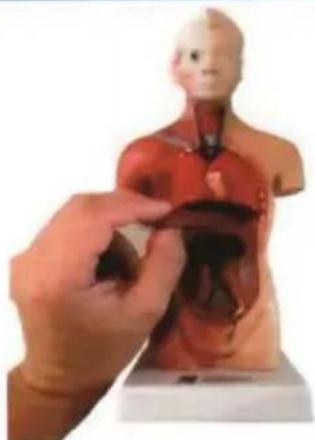
Hand holding a model of a human body in profile view (no text or symbols visible)- Add the Liver Right Underneath the Diaphragm
natural_image
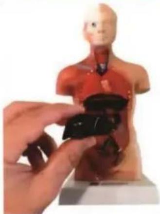
Hand holding a small black object next to a transparent model of a human torso (no text or symbols visible)- Add the Pancreas / Duodenum / Spleen to Upper Right Abdomen
natural_image
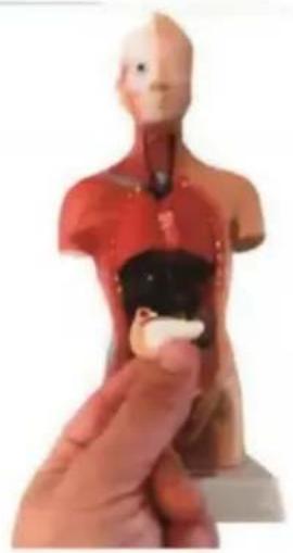
Illustration of a human figure with a hand holding an object, no visible text or symbols- CONT. Location of Pancreas / Duodenum/Spleen in Abdomen
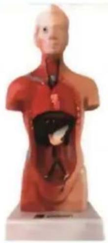
natural_image
Anatomical model of a human torso and arm, showing internal organs (no text or labels visible)- Add the Intestines and Stomach
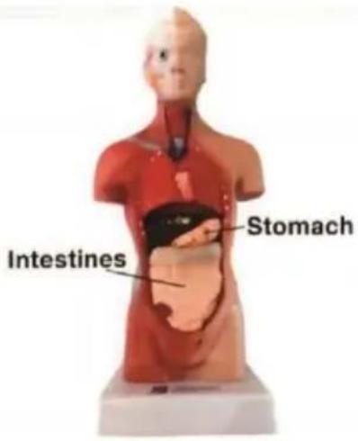
text_image
Intestines Stomach- Face the Torso Model Downward on a Flat Surface
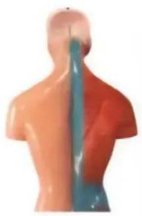
natural_image
Human back view showing a vertical spine with colored muscle fibers (no text or labels)- Add One Half(Hemisphere) of the Brain
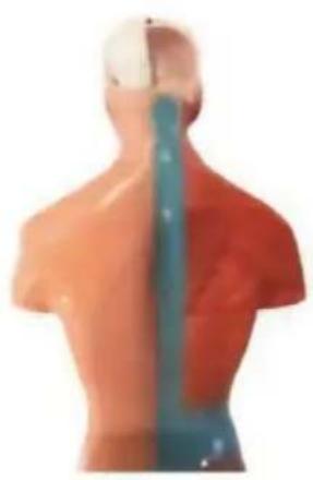
natural_image
Human back view showing a vertical spine with color gradient from orange to blue (no text or symbols)- Add the Other Half(Hemi-sphere) of the Brain
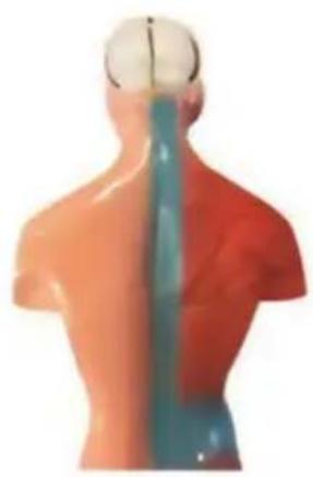
natural_image
Back view of a human torso with a vertical blue surgical spine (no text or symbols)- Add the Back of the Head
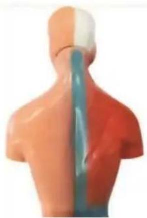
natural_image
Anatomical illustration of a human back and shoulder showing muscle fibers (no text or labels)
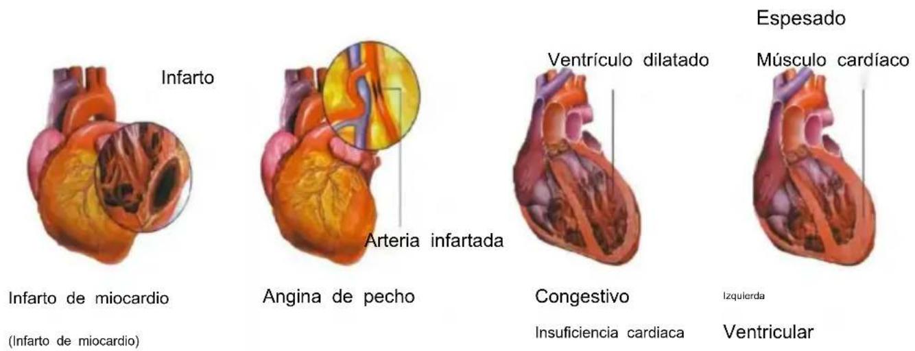
Hipertrofia
natural_image
Anatomical illustration of the human heart and major blood vessels (no text or labels)Aneurisma aórtico

text_image
TromboCerebrovascular
Accidente (derrame cerebral)
Aneurisma
Vasculitis
natural_image
Abstract illustration of a stylized figure with flowing hair against a red background (no text or symbols)- Ventrículo
natural_image
Close-up of a textured, irregularly shaped object with purple and gray sections (no visible text or symbols)- Lóbulo occipital Es
text_image
Left insula Frontal lobe Right insula Corpus striatum Corpus Callosum Internal Capsule Lentiform nucleus Brainstem (left half) Ventricles of brain Brainstem (Right half) Left Parietal lobe Right parietal lobe Cerebellum Temporal and occipital lobes with limbic system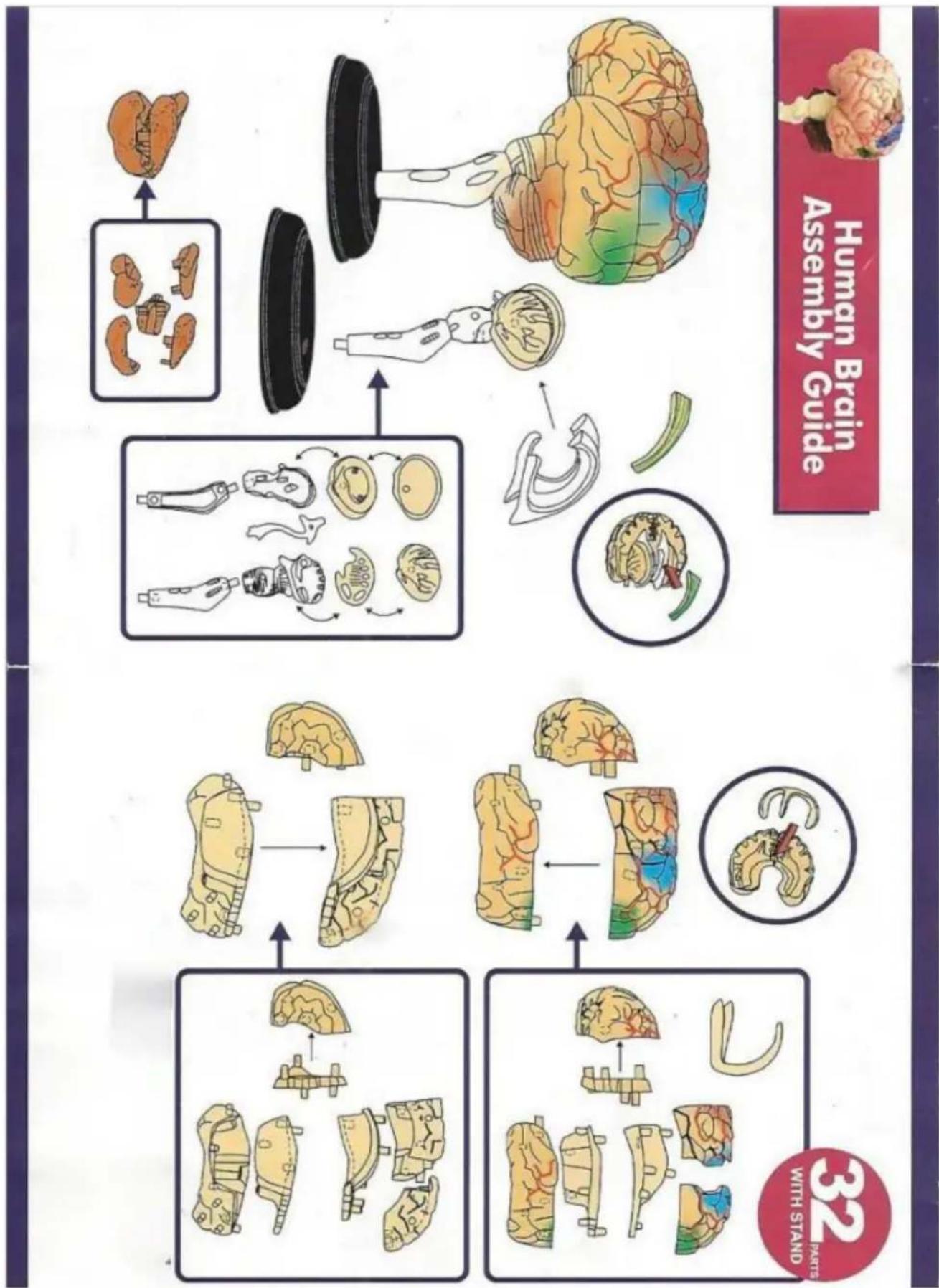
flowchart
graph TD
    A["Feeding"] --> B["Assembly"]
    B --> C["Brain with Human Brain"]
    C --> D["Assembly with Human Brain"]
    D --> E["Assembly with Human Brain"]
    E --> F["Assembly with Human Brain"]
    F --> G["Assembly with Human Brain"]
    G --> H["Assembly with Human Brain"]
    H --> I["Assembly with Human Brain"]
    I --> J["Assembly with Human Brain"]
    J --> K["Assembly with Human Brain"]
    K --> L["Assembly with Human Brain"]
    L --> M["Assembly with Human Brain"]
    M --> N["Assembly with Human Brain"]
    N --> O["Assembly with Human Brain"]
    O --> P["Assembly with Human Brain"]
    P --> Q["Assembly with Human Brain"]
    Q --> R["Assembly with Human Brain"]
    R --> S["Assembly with Human Brain"]
    S --> T["Assembly with Human Brain"]
    T --> U["Assembly with Human Brain"]
    U --> V["Assembly with Human Brain"]
    V --> W["Assembly with Human Brain"]
    W --> X["Assembly with Human Brain"]
    X --> Y["Assembly with Human Brain"]
    Y --> Z["Assembly with Human Brain"]
    Z --> AA["Assembly with Human Brain"]
    AA --> AB["Assembly with Human Brain"]
    AB --> AC["Assembly with Human Brain"]
    AC --> AD["Assembly with Human Brain"]
    AD --> AE["Assembly with Human Brain"]
    AE --> AF["Assembly with Human Brain"]
    AF --> AG["Assembly with Human Brain"]
    AG --> AH["Assembly with Human Brain"]
    AH --> AI["Assembly with Human Brain"]
    AI --> AJ["Assembly with Human Brain"]
    AJ --> AK["Assembly with Human Brain"]
    AK --> AL["Assembly with Human Brain"]
    AL --> AM["Assembly with Human Brain"]
    AM --> AN["Assembly with Human Brain"]
    AN --> AO["Assembly with Human Brain"]
    AO --> AP["Assembly with Human Brain"]
    AP --> AQ["Assembly with Human Brain"]
    AQ --> AR["Assembly with Human Brain"]
    AR --> AS["Assembly with Human Brain"]
    AS --> AT["Assembly with Human Brain"]
    AT --> AU["Assembly with Human Brain"]
    AU --> AV["Assembly with Human Brain"]
    AV --> AW["Assembly with Human Brain"]
    AW --> AX["Assembly with Human Brain"]
    AX --> AY["Assembly with Human Brain"]
Manufacturer: Shanghaimuxinmuyeyouxiangongsi Address: Shuangchenglu 803nong11hao1602A-1609shi, baoshanqu, shanghai 200000 CN. Imported to AUS: SIHAO PTY LTD. 1 ROKEVA STREET- EASTWOOD NSW 2122 Australia Imported to USA: Sanven Technology Ltd. Suite 250, 9166 Anaheim Place, Rancho Cucamonga, CA 91730

YH CONSULTING LIMITED. C/O YH Consulting Limited Office 147, Centurion House, London Road, Staines-upon-Thames, Surrey, TW18 4AX
VEVOR®
TOUGH TOOLS, HALF PRICE
natural_image
Medical illustrations of human anatomy including skeletal, skeletal muscle, internal organs, and brain models (no text or labels visible)POTRZEBUJESZ POMOCY? SKONTAKTUJ SIĘ Z NAMI!
natural_image
Simple line drawing of a human skeleton in profile view, no text or symbols presentAnatomia szkieletu
natural_image
Two identical white skeletal skeleton models displayed on pedestals, showing full skeletal structure and limbs (no text or labels visible)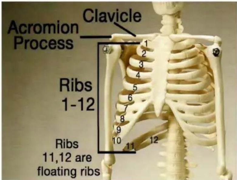
text_image
Acromion Process Clavicle Ribs 1-12 Ribs 11,12 are floating ribstext_image
Parietal Bone Frontal Bone Occipital Bone Sphenoid Bone Temporal Bone Maxilla Zygomatic Arch Mandibletext_image
Lamboid Suture Parietal Bone Parietal Bone Sagittal Suture Coronal Suture Frontal Bone- Lay the Torso Anatomy Model Flat on its Back on a Flat Surface
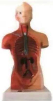
natural_image
Anatomical model of a human torso showing internal organs and structures (no text or labels visible)- Add the Posterior(Back)Half of the Right Lung to the Right Chest
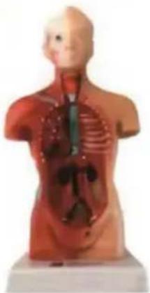
natural_image
Anatomical model of a human torso showing internal organs and structures (no text or labels visible)- Add the Posterior(Back)Half of the Left Lung to the Left Chest
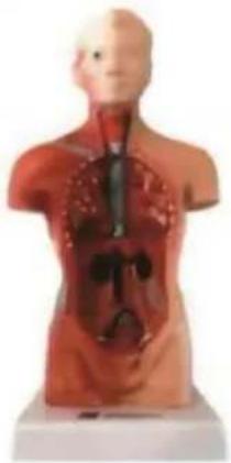
natural_image
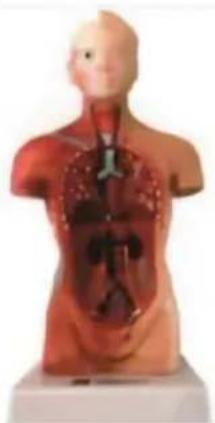
Anatomical model of a human torso showing internal organs (no text or labels visible)- Add the Trachea / Esophagus / Aorta
Piece Between the Lungs
natural_image
Anatomical model of a human torso and arm, showing internal organs (no text or labels visible)- Next Add The Heart on Top of the Trachea Piece
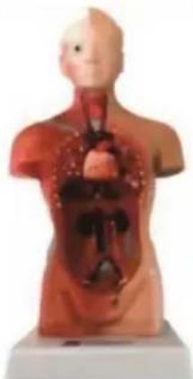
natural_image
Anatomical model of a human torso showing internal organs (no text or labels visible)- Add the Anterior(Front) Right Lung to the Right Cheae
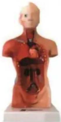
natural_image
Anatomical model of a human torso and arm, showing internal organs (no text or labels visible)- Add the Anterior (Front) Left Lung to the Left Chest
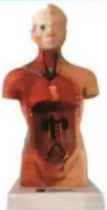
natural_image
Anatomical model of a human torso showing internal organs (no text or labels visible)- Add the Diaphragm Piece Right Underneath the Lungs
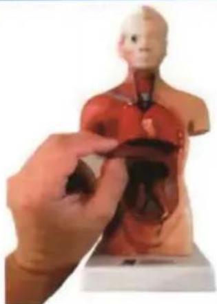
natural_image
Hand holding a model of a human body in profile view (no text or symbols visible)- Add the Liver Right Underneath the Diaphragm
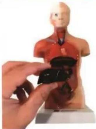
natural_image
Hand holding a small black object next to a transparent model of a human torso (no text or symbols visible)- Add the Pancreas / Duodenum / Spleen to Upper Right Abdomen
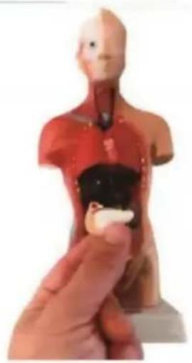
natural_image
Illustration of a human figure with a hand holding an object, no visible text or symbols- CONT. Location of Pancreas / Duodenum/Spleen in Abdomen
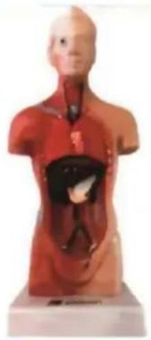
natural_image
Anatomical model of a human torso and arm, showing internal organs (no text or labels visible)- Add the Intestines and Stomach
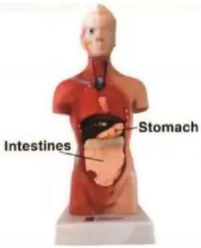
text_image
Intestines Stomach- Face the Torso Model Downward on a Flat Surface
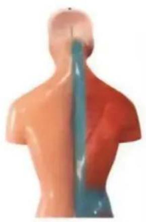
natural_image
Human back view showing a vertical spine split between orange and teal color (no text or symbols)- Add One Half(Hemisphere) of the Brain
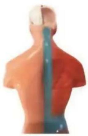
natural_image
Human back view showing a vertical spine with color gradient from orange to blue (no text or symbols)- Add the Other Half(Hemi-sphere) of the Brain
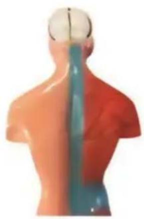
natural_image
Back view of a human torso with a vertical blue surgical band on the shoulder area (no text or symbols)- Add the Back of the Head
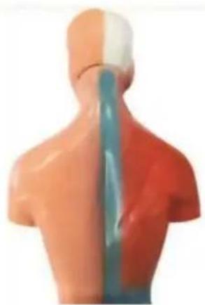
natural_image
Anatomical illustration of a human back and shoulder showing muscle fibers (no text or labels)
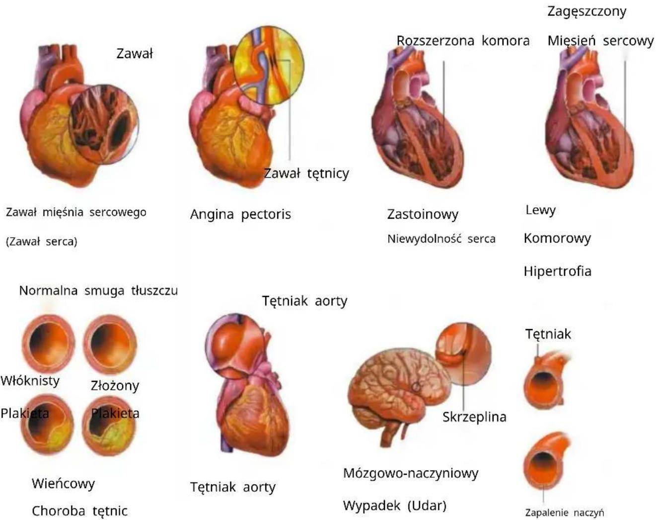
natural_image
Abstract illustration of a stylized figure with flowing hair against a red background (no text or symbols)- Komora
natural_image
Close-up of a textured, irregularly shaped object with a visible interior area (no text or symbols)text_image
Left insula Frontal lobe Right insula Corpus striatum Corpus Callosum Internal Capsule Lentiform nucleus Brainstem (left half) Ventricles of brain Brainstem (Right half) Left Parietal lobe Right parietal lobe Cerebellum Temporal and occipital lobes with limbic system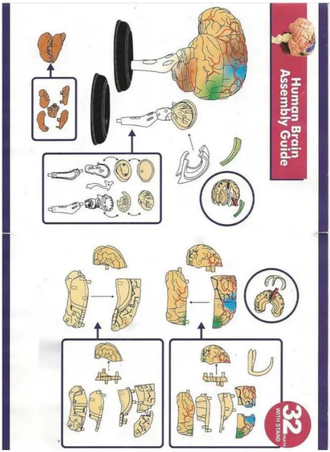
flowchart
graph TD
    A["Feeding"] --> B["Assembly"]
    B --> C["Brain Assembly"]
    C --> D["Human Brain Assembly Guide"]
    D --> E["Assembly"]
    E --> F["Final Assembly"]
    subgraph Human_Brain_1
        G["Red brain icon"] --> H["Human Brain icon"]
        I["Green brain icon"] --> J["Human Brain icon"]
        K["Blue brain icon"] --> L["Human Brain icon"]
        M["White brain icon"] --> N["Human Brain icon"]
        O["Black brain icon"] --> P["Human Brain icon"]
        Q["Yellow brain icon"] --> R["Human Brain icon"]
        S["Green brain icon"] --> T["Human Brain icon"]
        U["Blue brain icon"] --> V["Human Brain icon"]
        W["White brain icon"] --> X["Human Brain icon"]
        Y["Black brain icon"] --> Z["Human Brain icon"]
        AA["Yellow brain icon"] --> AB["Human Brain icon"]
        AC["Green brain icon"] --> AD["Human Brain icon"]
        AE["Blue brain icon"] --> AF["Human Brain icon"]
        AG["White brain icon"] --> AH["Human Brain icon"]
        AI["Black brain icon"] --> AJ["Human Brain icon"]
        AK["Yellow brain icon"] --> AL["Human Brain icon"]
        AM["Green brain icon"] --> AN["Human Brain icon"]
        AO["Blue brain icon"] --> AP["Human Brain icon"]
        AQ["White brain icon"] --> AR["Human Brain icon"]
        AS["Black brain icon"] --> AT["Human Brain icon"]
        AU["Yellow brain icon"] --> AV["Human Brain icon"]
        AW["Green brain icon"] --> AX["Human Brain icon"]
        AY["Red brain icon"] --> AZ["Human Brain Icon"]
    end
Manufacturer: Shanghaimuxinmuyeyouxiangongsi Address: Shuangchenglu 803nong11hao1602A-1609shi, baoshanqu, shanghai 200000 CN. Imported to AUS: SIHAO PTY LTD. 1 ROKEVA STREET- EASTWOOD NSW 2122 Australia Imported to USA: Sanven Technology Ltd. Suite 250, 9166 Anaheim Place, Rancho Cucamonga, CA 91730

YH CONSULTING LIMITED. C/O YH Consulting Limited Office 147, Centurion House, London Road, Staines-upon-Thames, Surrey, TW18 4AX
VEVOR®
TOUGH TOOLS, HALF PRICE
natural_image
Medical illustrations of human anatomy including skeletal, skeletal muscle, internal organs, and brain models (no text or labels visible)HULP NODIG? NEEM CONTACT MET ONS OP!
natural_image
Simple line drawing of a human skeleton in profile view, no text or symbols presentSkeletonatomie Het
natural_image
Two identical white skeletal models displayed on pedestals, showing full skeletal structure and limbs (no text or labels visible)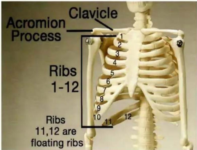
text_image
Acromion Process Clavicle Ribs 1-12 Ribs 11,12 are floating ribstext_image
Parietal Bone Frontal Bone Sphenoid Bone Occipital Bone Temporal Bone Maxilla Zygomatic Arch Mandibletext_image
Lamboid Suture Parietal Bone Parietal Bone Sagittal Suture Coronal Suture Frontal Bone- Lay the Torso Anatomy Model Flat on its Back on a Flat Surface
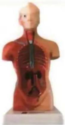
natural_image
Anatomical model of a human torso showing internal organs and structures (no text or labels visible)- Add the Posterior(Back)Half of the Right Lung to the Right Chest
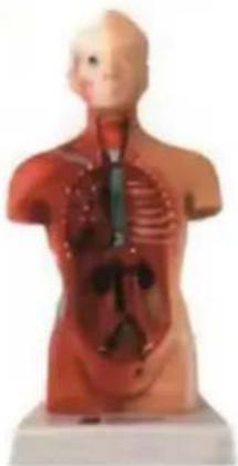
natural_image
Anatomical model of a human torso showing internal organs and structures (no text or labels visible)- Add the Posterior(Back)Half of the Left Lung to the Left Chest
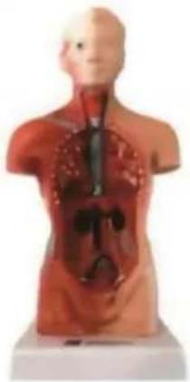
natural_image
Anatomical model of a human torso showing internal organs (no text or labels visible)- Add the Trachea / Esophagus / Aorta
Piece Between the Lungs
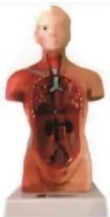
natural_image
Anatomical model of a human torso and arm, showing internal organs (no text or labels visible)- Next Add The Heart on Top of the Trachea Piece
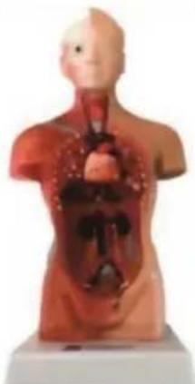
natural_image
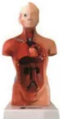
Anatomical model of a human torso showing internal organs (no text or labels visible)- Add the Anterior(Front) Right Lung to the Right Cheae
natural_image
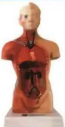
Anatomical model of a human torso and arm, showing internal organs (no text or labels visible)- Add the Anterior (Front) Left Lung to the Left Chest
natural_image
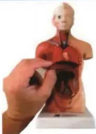
Anatomical model of a human torso showing internal organs (no text or labels visible)- Add the Diaphragm Piece Right Underneath the Lungs
natural_image
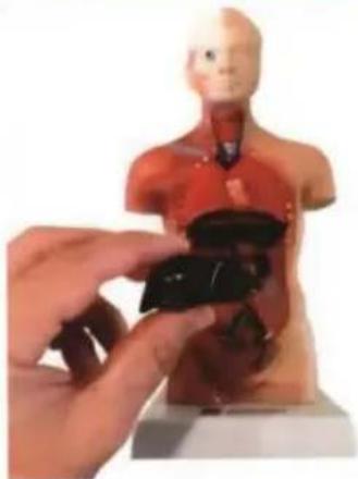
Hand holding a model of a human body in profile view (no text or symbols visible)- Add the Liver Right Underneath the Diaphragm
natural_image
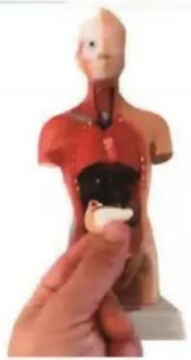
Hand holding a small black object next to a transparent model of a human torso (no text or symbols visible)- Add the Pancreas / Duodenum / Spleen to Upper Right Abdomen
natural_image
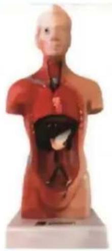
Illustration of a human figure with a hand holding an object, no visible text or symbols- CONT. Location of Pancreas / Duodenum/Spleen in Abdomen
natural_image
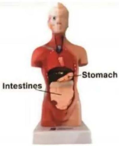
Anatomical model of a human torso and arm, showing internal organs (no text or labels visible)- Add the Intestines and Stomach
text_image
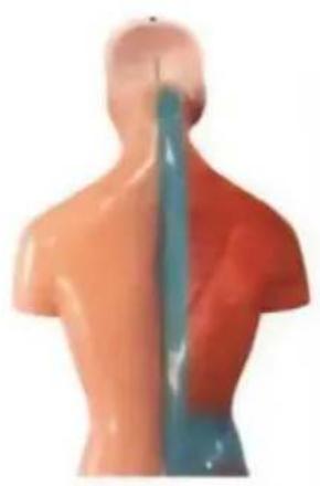
Intestines Stomach- Face the Torso Model Downward on a Flat Surface
natural_image
Human back view showing a vertical spine with colored muscle fibers (no text or labels)- Add One Half(Hemisphere) of the Brain
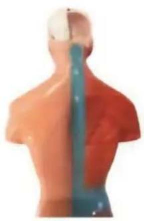
natural_image
Human back view showing a vertical spine with colored regions (no text or symbols)- Add the Other Half(Hemi-sphere) of the Brain
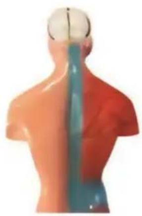
natural_image
Back view of a human torso with a vertical blue surgical band on the shoulder area (no text or symbols)- Add the Back of the Head
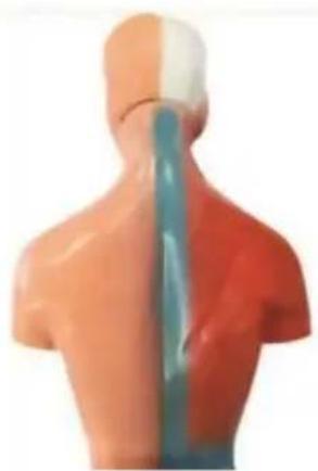
natural_image
Anatomical model of a human back showing internal muscle fibers (no text or labels)
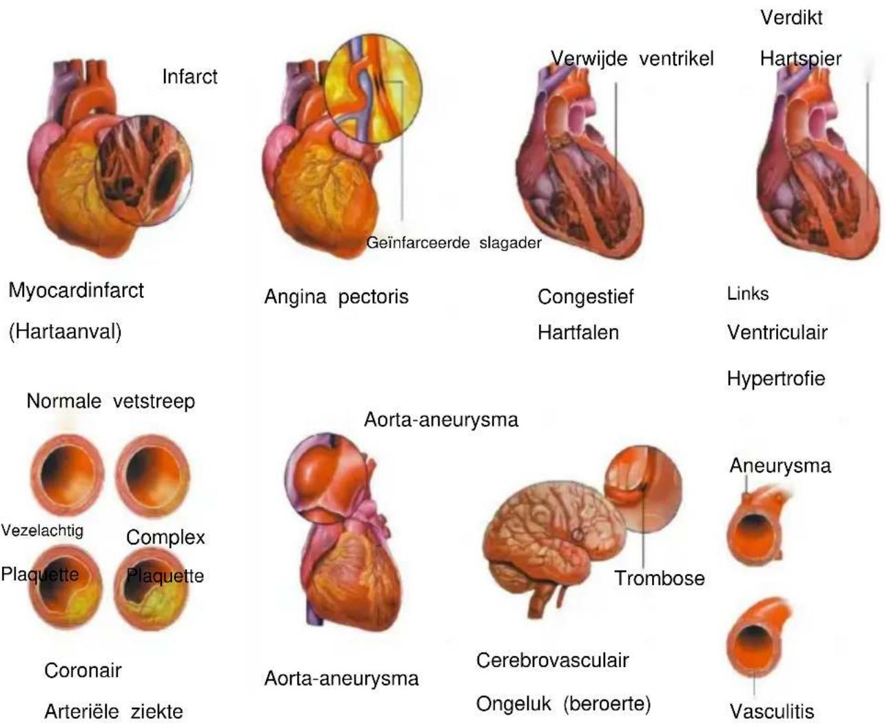
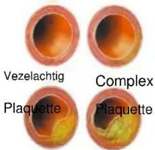
| 1. Rechter atrium2. Rechteroorschelp 3. Vena cava superior4. inferieure vena cava5. Rechterventrikel6. Longstam | 7. Opstijgende aorta8.Boog van de aorta9.Brachiocefale romp10.Gemeenschappelijke halsslagaderslagader 11.Arteriasubclavia 12.Arterieel ligament | 13. Afdalende aorta14. Linker oorschelp15. Linkerventrikel16. Rechterkransslagader17.Voorsteinterventriculaire tak vanlinker kransslagader |
| 18.Circumflexe tak vanlinker kransslagader 19.Grote hartader20.Middenhartader21. Kleine hartader22.Achtersteinterventriculaire tak vanrechter kransslagader | 23.Coronaire sinus24. Fossa ovalis25.Opening van de kransslagadersinus26.Tricuspidalisklep27.Pulmonalisklep28.Linker atrium | 29.Bicuspide klep (mitralisklep)30.Aortaklep31. Linker longslagader 32.Linker longader 33. Rechterlongslagader 34. Rechterlongader |
1. Frontale kwab
natural_image
Abstract illustration of a stylized figure with flowing hair against a red background (no text or symbols)5.Ventrikel
natural_image
Close-up of a textured, irregularly shaped object with a visible interior area (no text or symbols)6.Occipitale kwab Dit
text_image
Left insula Frontal lobe Right insula Corpus striatum Corpus Callosum Internal Capsule Lentiform nucleus Brainstem (left half) Ventricles of brain Brainstem (Right half) Left Parietal lobe Right parietal lobe Cerebellum Temporal and occipital lobes with limbic system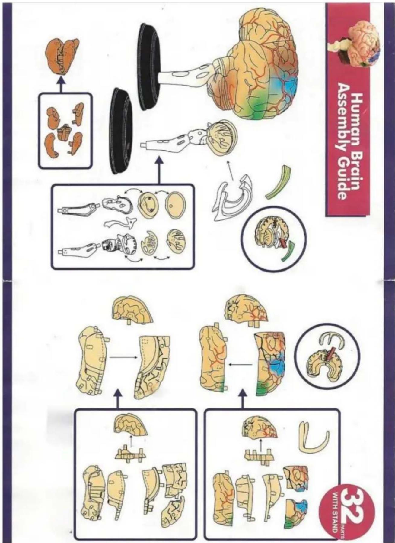
flowchart
graph TD
    A["Feeding"] --> B["Assembly"]
    B --> C["Brain with Human Brain"]
    C --> D["Assembly with Human Brain"]
    D --> E["Assembly with Human Brain"]
    E --> F["Assembly with Human Brain"]
    F --> G["Assembly with Human Brain"]
    G --> H["Assembly with Human Brain"]
    H --> I["Assembly with Human Brain"]
    I --> J["Assembly with Human Brain"]
    J --> K["Assembly with Human Brain"]
    K --> L["Assembly with Human Brain"]
    L --> M["Assembly with Human Brain"]
    M --> N["Assembly with Human Brain"]
    N --> O["Assembly with Human Brain"]
    O --> P["Assembly with Human Brain"]
    P --> Q["Assembly with Human Brain"]
    Q --> R["Assembly with Human Brain"]
    R --> S["Assembly with Human Brain"]
    S --> T["Assembly with Human Brain"]
    T --> U["Assembly with Human Brain"]
    U --> V["Assembly with Human Brain"]
    V --> W["Assembly with Human Brain"]
    W --> X["Assembly with Human Brain"]
    X --> Y["Assembly with Human Brain"]
    Y --> Z["Assembly with Human Brain"]
    Z --> AA["Assembly with Human Brain"]
    AA --> AB["Assembly with Human Brain"]
    AB --> AC["Assembly with Human Brain"]
    AC --> AD["Assembly with Human Brain"]
    AD --> AE["Assembly with Human Brain"]
    AE --> AF["Assembly with Human Brain"]
    AF --> AG["Assembly with Human Brain"]
    AG --> AH["Assembly with Human Brain"]
    AH --> AI["Assembly with Human Brain"]
    AI --> AJ["Assembly with Human Brain"]
    AJ --> AK["Assembly with Human Brain"]
    AK --> AL["Assembly with Human Brain"]
    AL --> AM["Assembly with Human Brain"]
    AM --> AN["Assembly with Human Brain"]
    AN --> AO["Assembly with Human Brain"]
    AO --> AP["Assembly with Human Brain"]
    AP --> AQ["Assembly with Human Brain"]
    AQ --> AR["Assembly with Human Brain"]
    AR --> AS["Assembly with Human Brain"]
    AS --> AT["Assembly with Human Brain"]
    AT --> AU["Assembly with Human Brain"]
    AU --> AV["Assembly with Human Brain"]
    AV --> AW["Assembly with Human Brain"]
    AW --> AX["Assembly with Human Brain"]
    AX --> AY["Assembly with Human Brain"]
Manufacturer: Shanghaimuxinmuyeyouxiangongsi Address: Shuangchenglu 803nong11hao1602A-1609shi, baoshanqu, shanghai 200000 CN. Imported to AUS: SIHAO PTY LTD. 1 ROKEVA STREET- EASTWOOD NSW 2122 Australia Imported to USA: Sanven Technology Ltd. Suite 250, 9166 Anaheim Place, Rancho Cucamonga, CA 91730

YH CONSULTING LIMITED. C/O YH Consulting Limited Office 147, Centurion House, London Road, Staines-upon-Thames, Surrey, TW18 4AX
VEVOR®
TOUGH TOOLS, HALF PRICE
garantiecertificaat www.vevor.com/support
VEVOR®
TOUGH TOOLS, HALF PRICE
natural_image
Medical illustrations of human anatomy including skeletal, skeletal muscle, internal organs, and cerebral blood vessels (no text or labels visible)BEHÖVER HJÄLP? KONTAKTA OSS!
natural_image
Simple line drawing of a human skeleton in profile view, no text or symbols presentSkelettanatomi
natural_image
Two identical white skeletal skeleton models displayed on pedestals, showing full skeletal structure and limbs (no text or labels visible)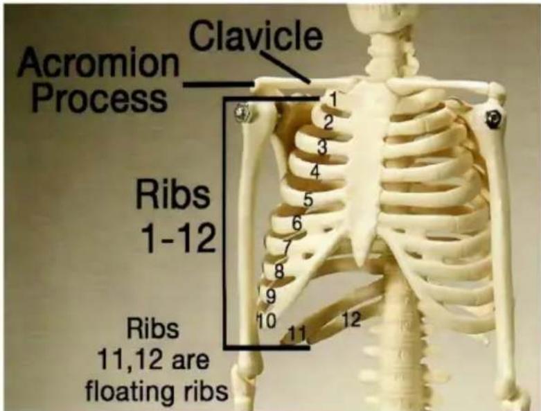
text_image
Acromion Process Clavicle Ribs 1-12 Ribs 11,12 are floating ribstext_image
Parietal Bone Frontal Bone Occipital Bone Sphenoid Bone Temporal Bone Maxilla Zygomatic Arch Mandibletext_image
Lamboid Suture Parietal Bone Parietal Bone Sagittal Suture Coronal Suture Frontal Bone- Lay the Torso Anatomy Model Flat on its Back on a Flat Surface
natural_image
Anatomical model of a human torso showing internal organs and structures (no text or labels visible)- Add the Posterior(Back)Half of the Right Lung to the Right Chest
natural_image
Anatomical model of a human torso showing internal organs and structures (no text or labels visible)- Add the Posterior(Back)Half of the Left Lung to the Left Chest
natural_image
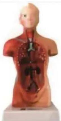
Anatomical model of a human torso showing internal organs (no text or labels visible)- Add the Trachea / Esophagus / Aorta
Piece Between the Lungs
natural_image
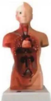
Anatomical model of a human torso and arm, showing internal organs (no text or labels visible)- Next Add The Heart on Top of the Trachea Piece
natural_image
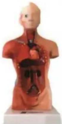
Anatomical model of a human torso showing internal organs (no text or labels visible)- Add the Anterior(Front) Right Lung to the Right Cheae
natural_image
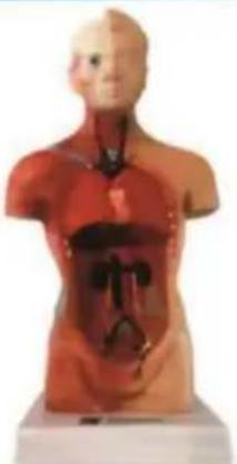
Anatomical model of a human torso and arm, showing internal organs (no text or labels visible)- Add the Anterior (Front) Left Lung to the Left Chest
natural_image
Anatomical model of a human torso showing internal organs (no text or labels visible)- Add the Diaphragm Piece Right Underneath the Lungs
natural_image
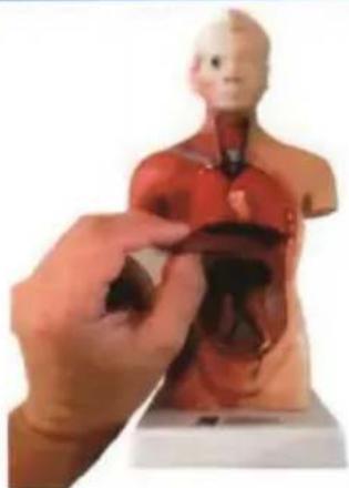
Hand holding a model of a human body in profile view (no text or symbols visible)- Add the Liver Right Underneath the Diaphragm
natural_image
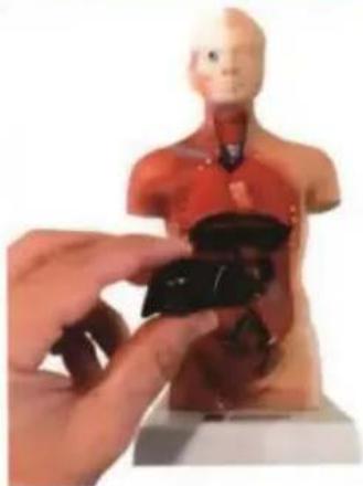
Hand holding a small black object next to a transparent model of a human torso (no text or symbols visible)- Add the Pancreas / Duodenum / Spleen to Upper Right Abdomen
natural_image
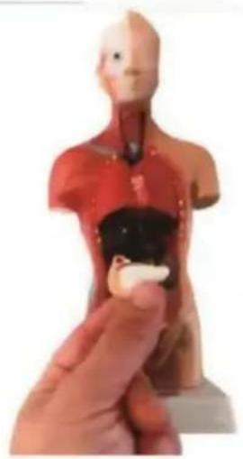
Illustration of a human figure with a hand holding an object, no visible text or symbols- CONT. Location of Pancreas / Duodenum/Spleen in Abdomen
natural_image
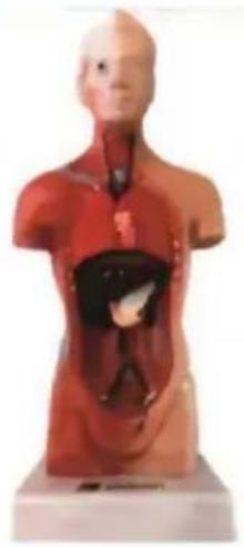
Anatomical model of a human torso and arm, showing internal organs (no text or labels visible)- Add the Intestines and Stomach
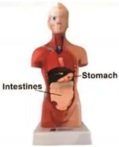
text_image
Intestines Stomach- Face the Torso Model Downward on a Flat Surface
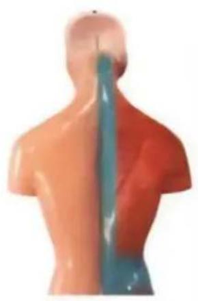
natural_image
Human back view showing a vertical spine with colored muscle fibers (no text or labels)- Add One Half(Hemisphere) of the Brain
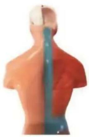
natural_image
Human back view showing a vertical spine with colored regions (no text or symbols)- Add the Other Half(Hemi-sphere) of the Brain
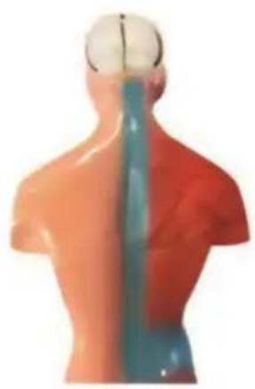
natural_image
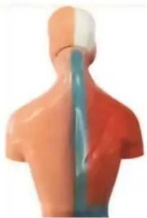
Back view of a human torso with a vertical blue surgical band on the shoulder area (no text or symbols)- Add the Back of the Head
natural_image
Anatomical model of a human back showing internal muscle structure (no text or labels)
natural_image
Abstract illustration of a stylized figure with flowing hair against a red background (no text or symbols)5. Ventrikel
natural_image
Close-up of a textured, irregularly shaped object with purple and gray sections (no visible text or symbols)6. Occipitallob
text_image
Left insula Frontal lobe Right insula Corpus striatum Corpus Callosum Internal Capsule Lentiform nucleus Brainstem (left half) Ventricles of brain Brainstem (Right half) Left Parietal lobe Right parietal lobe Cerebellum Temporal and occipital lobes with limbic system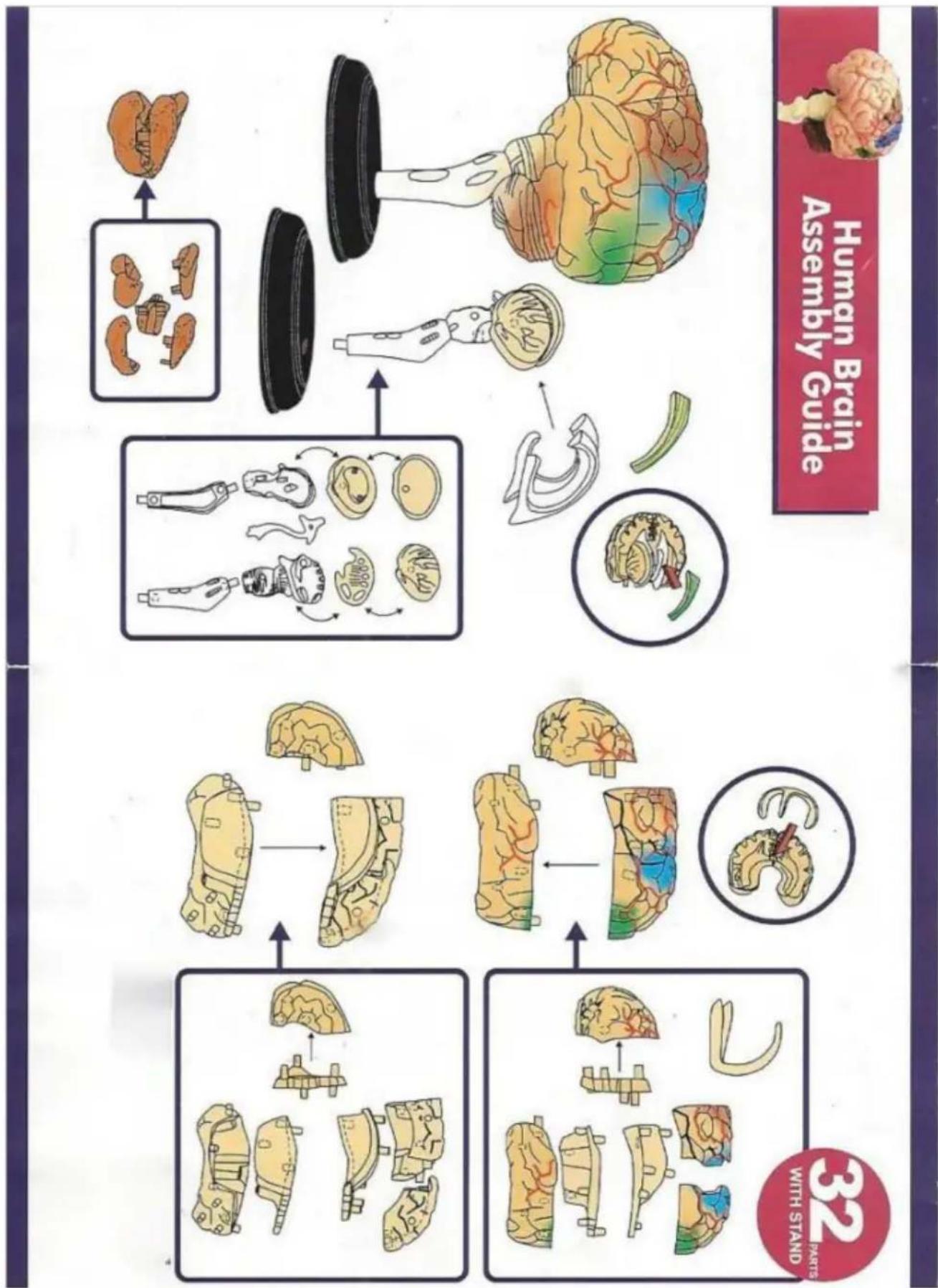
flowchart
graph TD
    A["Feeding"] --> B["Assembly"]
    B --> C["Brain with Human Brain"]
    C --> D["Assembly with Human Brain"]
    D --> E["Assembly with Human Brain"]
    E --> F["Assembly with Human Brain"]
    F --> G["Assembly with Human Brain"]
    G --> H["Assembly with Human Brain"]
    H --> I["Assembly with Human Brain"]
    I --> J["Assembly with Human Brain"]
    J --> K["Assembly with Human Brain"]
    K --> L["Assembly with Human Brain"]
    L --> M["Assembly with Human Brain"]
    M --> N["Assembly with Human Brain"]
    N --> O["Assembly with Human Brain"]
    O --> P["Assembly with Human Brain"]
    P --> Q["Assembly with Human Brain"]
    Q --> R["Assembly with Human Brain"]
    R --> S["Assembly with Human Brain"]
    S --> T["Assembly with Human Brain"]
    T --> U["Assembly with Human Brain"]
    U --> V["Assembly with Human Brain"]
    V --> W["Assembly with Human Brain"]
    W --> X["Assembly with Human Brain"]
    X --> Y["Assembly with Human Brain"]
    Y --> Z["Assembly with Human Brain"]
    Z --> AA["Assembly with Human Brain"]
    AA --> AB["Assembly with Human Brain"]
    AB --> AC["Assembly with Human Brain"]
    AC --> AD["Assembly with Human Brain"]
    AD --> AE["Assembly with Human Brain"]
    AE --> AF["Assembly with Human Brain"]
    AF --> AG["Assembly with Human Brain"]
    AG --> AH["Assembly with Human Brain"]
    AH --> AI["Assembly with Human Brain"]
    AI --> AJ["Assembly with Human Brain"]
    AJ --> AK["Assembly with Human Brain"]
    AK --> AL["Assembly with Human Brain"]
    AL --> AM["Assembly with Human Brain"]
    AM --> AN["Assembly with Human Brain"]
    AN --> AO["Assembly with Human Brain"]
    AO --> AP["Assembly with Human Brain"]
    AP --> AQ["Assembly with Human Brain"]
    AQ --> AR["Assembly with Human Brain"]
    AR --> AS["Assembly with Human Brain"]
    AS --> AT["Assembly with Human Brain"]
    AT --> AU["Assembly with Human Brain"]
    AU --> AV["Assembly with Human Brain"]
    AV --> AW["Assembly with Human Brain"]
    AW --> AX["Assembly with Human Brain"]
    AX --> AY["Assembly with Human Brain"]
Manufacturer: Shanghaimuxinmuyeyouxiangongsi Address: Shuangchenglu 803nong11hao1602A-1609shi, baoshanqu, shanghai 200000 CN. Imported to AUS: SIHAO PTY LTD. 1 ROKEVA STREET- EASTWOOD NSW 2122 Australia Imported to USA: Sanven Technology Ltd. Suite 250, 9166 Anaheim Place, Rancho Cucamonga, CA 91730

YH CONSULTING LIMITED. C/O YH Consulting Limited Office 147, Centurion House, London Road, Staines-upon-Thames, Surrey, TW18 4AX
VEVOR®
TOUGH TOOLS, HALF PRICE
www.vevor.com/support