LDX-V4 - Vevor - Free user manual and instructions
Find the device manual for free LDX-V4 Vevor in PDF.


User questions about LDX-V4 Vevor
0 question about this device. Answer the ones you know or ask your own.
Ask a new question about this device
Download the instructions for your in PDF format for free! Find your manual LDX-V4 - Vevor and take your electronic device back in hand. On this page are published all the documents necessary for the use of your device. LDX-V4 by Vevor.
USER MANUAL LDX-V4 Vevor
Technical Support and E-Warranty Certificate www.vevor.com/support
HUMAN BODY TEACHING MODELS USER MANUAL
MODEL:LDX-V4
We continue to be committed to provide you tools with competitive price. "Save Half", "Half Price" or any other similar expressions used by us only represents an estimate of savings you might benefit from buying certain tools with us compared to the major top brands and does not necessarily mean to cover all categories of tools offered by us. You are kindly reminded to verify carefully when you are placing an order with us if you are actually saving half in comparison with the top major brands
VEVOR®
TOUGH TOOLS, HALF PRICE
HUMAN BODY TEACHING
MODELS
MODEL: LDX-V4
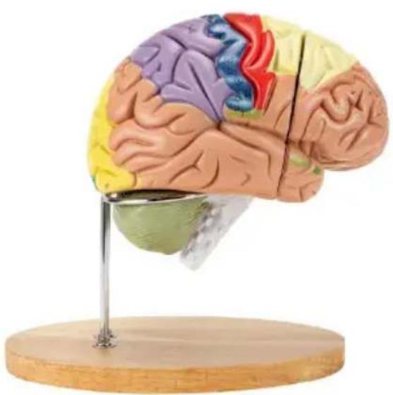
natural_image
Illustration of a human brain model with colored cortical areas and a metallic stand (no text or labels)NEED HELP? CONTACT US!
Have product questions? Need technical support? Please feel fr contact us:
Technical Support and E-Warranty Certificate www.vevor.com/support
This is the original instruction, please read all manual instructions carefully before operating. VEVOR reserves a clear interpretation of user manual. The appearance of the product shall be subject to product you received. Please forgive us that we won't inform your use, there are any technology or software updates on our product.
SAFETY INSTRUCTIONS
WARNING:
- Read this instructions before using this product. Failure to do so can result in serious injury.
- After use, please assemble all the parts and keep them vertically.
- Avoid high temperature and direct sunlight.
- Remove minor stains with neutral detergent. The use of alcohol, beer and other substances may damage the product.
SAVE THESE INSTRUCTIONS
PRODUCT PARAMETER
| Model | LDX-V4 |
| Labeling Mode | Color And Number |
| Material | PVC |
| Removable Parts | 4 |
2 times magnification anatomical model of human brain
Left Cerebral Hemisphere-Lateral View
- Motor area(somatomotor cortex)
1.Sensory area (somatosensory cortex)
2.Frontal lobe
3.Temporal lobe
4.Parietal lobe
5.Occipital lobe
6.Left Hemisphere of cerebellum
7.Superior frontal gyrus
8.Middle frontal gyrus
9.Inferior frontal gyrus
10.Precentral gyrus(somatomotor cortex)
11.Postcentral gyrus(somatosensory cortex)
12.Superior parietal lobule
13.Inferior parietal lobule 3
14.Lateral occipital gyrus
15.Superior temporal gyrus
16.Middle temporal gyrus
17.Inferior temporal gyrus
18.Superior frontal sulcus
19.Pre-central sulcus
20.Central sulcus
21.Post-central sulcus
22.Intraparietal sulcus
23.Transverse occipital sulcus
24.Lateral occipital sulcus
25.Parieto-occipital sulcus
26.Superior temporal sulcus
27.Inferior temporal sulcus
28.Lateral cerebral sulcus(Sylvian fissure)
29.Anterior horizontal ramus of Sylvian fissure
30.Anterior ascending ramus of Sylvian fissure
- Posterior ramus of Sylvian fissure
32.Inferior temporal gyrus
33.Paracentral lobule
34.Precuneus
35.Cuneus
36.Calcarine sulcus
37.Cingulate gyrus
38.Cingulate sulcus
39.Subfrontal part of cingulate sulcus
40.Sulcus of corpus callosum
41.Trunk of corpus callosum
42.Genu of corpus callosum
43.Splenium of corpus callosum
44.Septum pellucidum
45.Body of fornix
46.Anterior commissure
47.Interthalamic adhesion(Intermediate mass)
- Posterior commissure
49.Thalamus
50.Interventricular foramen(Monro)
51.Hypothalamic sulcus
52.Lamina terminalis
53.Optic chiasm
54.Pineal body
55.Pineal recess
56.Corpora quadrigemina
57.Cerebral aqueduct(Sylvius)
58.Superior medullary velum
59.Fourth ventricle
60.Central canal of medulla oblongata
61.Arbor vitae
62.Vermis of cerebellum
63.Medulla oblongata
64.Medial longitudinal fasciculus
65.Pons
66.Longitudinal fibers of pons
67.Cerebral peduncle
68.Tuber cinereum
69.Mammillary body
70.Interpeduncular recess
- Optic recess
72 Infundibular recess
Right Cerebral Hemisphere
73.Basal surface
74.Orbital gyri
75.Straight gyrus
76.Fusiform gyrus(Lateral occipitotemporal gyrus)
77.Lingual gyrus
78.Hippocampal gyrus
79.Uncus
- Internal capsule
81.External capsule
82.Corona radiata
83.Inferior surface of corpus callosum
84.Short gyri of insula
85.Fibers of corpus
86.insula
87.Claustrum
88.Lateral ventricle(anterior horn)
89.Lateral ventricle (posterior horn)
90.Fornix
91.Anterior perforated substance
- Olfactory tract(Cranial nerve I
93.Lateral olfactory stria
94.Medial olfactory stria
Right Brain Stem and Cerebellum
- Branches of internal carotid arterio vena terminalis
96 Lentiform nucleus
97 Corona radiata
98 Caudate nucleus
99.5.1 terminals
- Superior colliculi of corpora quadrigemina
103.Lateral geniculate body
104.Lateral sulcus of mesencephalon
105.Trigonum lemniscus
106.Frenulum of superior medullary vellum
107.Superior quadrigeminal brachium
108.Inferior quadrigeminal brachium
109.Pulvinar of thalamus
- Posterior perforated substance
111.Optic tract
112.Optic nerve (Cranial nerve II)
113.Cavity of third ventricle
114.Oculomotor nerve(Cranial nerve III)
115.Pituitary gland
116.Trochlear nerve(Cranial nerve IV)
117.Trigeminal nerve(Cranial nerve V)
118.Abducent nerve(Cranial nerve VI)
119.Facial nerve(Cranial nerve VII)
120.Vestibulocochlear nerve(Cranial nerve VIII)
121.Glossopharyngeal nerve (Cranial nerve IX)
122.Vagus nerve(Cranial nerve X)
123.Accessory nerve(Cranial nerve XI)
124.Hypoglossal nerve(Cranial nerve XII)
125.Olive
126.Pyramidal tracts
127.Decussation of pyramids
128.Flocculus of cerebellum
129.Tonsil of cerebellum
130 Vermis of cerebellum
Manufacturer: Shanghaimuxinmuyeyouxiangongsi
Address: Shuangchenglu 803nong11hao1602A-1609shi, baoshanqu, shanghai 200000 CN.
Imported to AUS: SIHAO PTY LTD. 1 ROKEVA STREET-EASTWOOD NSW 2122 Australia
Imported to USA: Sanven Technology Ltd. Suite 250, 9166 Anaheim Place, Rancho Cucamonga, CA 91730
| EC | REP |
YH CONSULTING LIMITED. C/O YH Consulting Limited Office 147, Centurion House, London Road, Staines-upon-Thames, Surrey, TW18 4AX
VEVOR®
TOUGH TOOLS, HALF PRICE
Technical Support and E-Warranty Certificate
www.vevor.com/support
VEVOR®
TOUGH TOOLS, HALF PRICE
natural_image
Anatomical model of a human brain with colored cortical areas, mounted on a wooden stand (no text or labels visible)BESOIN D'AIDE? CONTACTEZ-NOUS!
2 times magnification anatomical model of human brain
Left Cerebral Hemisphere-Lateral View
1.Motor area(somatomotor cortex)
1.Sensory area (somatosensory cortex)
2.Frontal lobe
- Temporal lobe
4.Parietal lobe
5.Occipital lobe
6.Left Hemisphere of cerebellum
7.Superior frontal gyrus
8.Middle frontal gyrus
9.Inferior frontal gyrus
10.Precentral gyrus(somatomotor cortex)
11.Postcentral gyrus(somatosensory cortex)
12.Superior parietal lobule
13.Inferior parietal lobule 3
14.Lateral occipital gyrus
15.Superior temporal gyrus
16.Middle temporal gyrus
17.Inferior temporal gyrus
18.Superior frontal sulcus
19.Pre-central sulcus
20.Central sulcus
21.Post-central sulcus
22.Intraparietal sulcus
23.Transverse occipital sulcus
24.Lateral occipital sulcus
25.Parieto-occipital sulcus
26.Superior temporal sulcus
27.Inferior temporal sulcus
28.Lateral cerebral sulcus(Sylvian fissure)
29.Anterior horizontal ramus of Sylvian fissure
30.Anterior ascending ramus of Sylvian fissure
- Posterior ramus of Sylvian fissure
32.Inferior temporal gyrus
33.Paracentral lobule
34.Precuneus
35.Cuneus
36.Calcarine sulcus
37.Cingulate gyrus
38.Cingulate sulcus
39.Subfrontal part of cingulate sulcus
40.Sulcus of corpus callosum
41.Trunk of corpus callosum
42.Genu of corpus callosum
43.Splenium of corpus callosum
44.Septum pellucidum
45.Body of fornix
46.Anterior commissure
47.Interthalamic adhesion(Intermediate mass)
- Posterior commissure
49.Thalamus
50.Interventricular foramen(Monro)
51.Hypothalamic sulcus
52.Lamina terminalis
53.Optic chiasm
54.Pineal body
55.Pineal recess
56.Corpora quadrigemina
57.Cerebral aqueduct(Sylvius)
58.Superior medullary velum
59.Fourth ventricle
60.Central canal of medulla oblongata
61.Arbor vitae
62.Vermis of cerebellum
63.Medulla oblongata
64.Medial longitudinal fasciculus
65.Pons
66.Longitudinal fibers of pons
67.Cerebral peduncle
68.Tuber cinereum
69.Mammillary body
70.Interpeduncular recess
71.Optic recess
72 Infundibular recess
Right Cerebral Hemisphere
73.Basal surface
74.Orbital gyri
75.Straight gyrus
76.Fusiform gyrus(Lateral occipitotemporal gyrus)
77.Lingual gyrus
78.Hippocampal gyrus
79.Uncus
- Internal capsule
81.External capsule
82.Corona radiata
83.Inferior surface of corpus callosum
84.Short gyri of insula
85.Fibers of corpus
86.insula
87.Claustrum
88.Lateral ventricle(anterior horn)
89.Lateral ventricle (posterior horn)
90.Fornix
- Anterior perforated substance
92 Olfactory tract(Cranial nerve 1
93.Lateral olfactory stria
94.Medial olfactory stria
Right Brain Stem and Cerebellum
95 Branches of internal carotid artery vena terminalis
96Lentiform nucleus
97 Corona radiata
98 Caudate nucleus
99.Stuy terminals
-
Superior coliculi of corpora quadrigemina
-
Infering coilrculi of corpora quadrigemina
102.Medial gemiculate body
103.Lateral geniculate body
104.Lateral sulcus of mesencephalon
105.Trigonum lemniscus
- Frenulum of superior medullary vellum
107.Superior quadrigeminal brachium
108.Inferior quadrigeminal brachium
109.Pulvinar of thalamus
- Posterior perforated substance
111.Optic tract
112.Optic nerve (Cranial nerve II)
113.Cavity of third ventricle
114.Oculomotor nerve(Cranial nerve III)
115.Pituitary gland
116.Trochlear nerve(Cranial nerve IV)
117.Trigeminal nerve(Cranial nerve V)
118.Abducent nerve(Cranial nerve VI)
119.Facial nerve(Cranial nerve VII)
120.Vestibulocochlear nerve(Cranial nerve VIII)
121.Glossopharyngeal nerve (Cranial nerve IX)
122.Vagus nerve(Cranial nerve X)
123.Accessory nerve(Cranial nerve XI)
124.Hypoglossal nerve(Cranial nerve XII)
125.Olive
126.Pyramidal tracts
127.Decussation of pyramids
128.Flocculus of cerebellum
129.Tonsil of cerebellum
130 Vermis of cerebellum
Manufacturer: Shanghaimuxinmuyeyouxiangongsi
Address: Shuangchenglu 803nong11hao1602A-1609shi, baoshanqu, shanghai 200000 CN.
Imported to AUS: SIHAO PTY LTD. 1 ROKEVA STREET-EASTWOOD NSW 2122 Australia
Imported to USA: Sanven Technology Ltd. Suite 250, 9166 Anaheim Place, Rancho Cucamonga, CA 91730
| EC | REP |
YH CONSULTING LIMITED. C/O YH Consulting Limited Office 147, Centurion House, London Road, Staines-upon-Thames, Surrey, TW18 4AX
VEVOR®
TOUGH TOOLS, HALF PRICE
natural_image
Anatomical model of a human brain with colored cortical areas, mounted on a wooden stand (no text or labels visible)2 times magnification anatomical model of human brain
Left Cerebral Hemisphere-Lateral View
- Motor area(somatomotor cortex)
1.Sensory area (somatosensory cortex)
2.Frontal lobe
- Temporal lobe
4.Parietal lobe
5.Occipital lobe
6.Left Hemisphere of cerebellum
7.Superior frontal gyrus
8.Middle frontal gyrus
9.Inferior frontal gyrus
10.Precentral gyrus(somatomotor cortex)
11.Postcentral gyrus(somatosensory cortex)
12.Superior parietal lobule
13.Inferior parietal lobule 3
14.Lateral occipital gyrus
15.Superior temporal gyrus
16.Middle temporal gyrus
17.Inferior temporal gyrus
18.Superior frontal sulcus
19.Pre-central sulcus
20.Central sulcus
21.Post-central sulcus
22.Intraparietal sulcus
23.Transverse occipital sulcus
24.Lateral occipital sulcus
25.Parieto-occipital sulcus
26.Superior temporal sulcus
27.Inferior temporal sulcus
28.Lateral cerebral sulcus(Sylvian fissure)
29.Anterior horizontal ramus of Sylvian fissure
30.Anterior ascending ramus of Sylvian fissure
- Posterior ramus of Sylvian fissure
32.Inferior temporal gyrus
33.Paracentral lobule
34.Precuneus
35.Cuneus
36.Calcarine sulcus
37.Cingulate gyrus
38.Cingulate sulcus
39.Subfrontal part of cingulate sulcus
40.Sulcus of corpus callosum
41.Trunk of corpus callosum
42.Genu of corpus callosum
43.Splenium of corpus callosum
44.Septum pellucidum
45.Body of fornix
46.Anterior commissure
47.Interthalamic adhesion(Intermediate mass)
- Posterior commissure
49.Thalamus
50.Interventricular foramen(Monro)
51.Hypothalamic sulcus
52.Lamina terminalis
53.Optic chiasm
54.Pineal body
55.Pineal recess
56.Corpora quadrigemina
57.Cerebral aqueduct(Sylvius)
58.Superior medullary velum
59.Fourth ventricle
60.Central canal of medulla oblongata
61.Arbor vitae
62.Vermis of cerebellum
63.Medulla oblongata
64.Medial longitudinal fasciculus
65.Pons
66.Longitudinal fibers of pons
67.Cerebral peduncle
68.Tuber cinereum
69.Mammillary body
70.Interpeduncular recess
71.Optic recess
72 Infundibular recess
Right Cerebral Hemisphere
73.Basal surface
74.Orbital gyri
75.Straight gyrus
76.Fusiform gyrus(Lateral occipitotemporal gyrus)
77.Lingual gyrus
78.Hippocampal gyrus
79.Uncus
- Internal capsule
81.External capsule
82.Corona radiata
83.Inferior surface of corpus callosum
84.Short gyri of insula
85.Fibers of corpus
86.insula
87.Claustrum
88.Lateral ventricle(anterior horn)
89.Lateral ventricle (posterior horn)
90.Fornix
- Anterior perforated substance
92 Olfactory tract(Cranial nerve 1
93.Lateral olfactory stria
94.Medial olfactory stria
Right Brain Stem and Cerebellum
95 Branches of internal carotid artery & vena terminalis
96Lentiform nucleus
97 Corona radiata
98 Caudate nucleus
-
St. terminals
-
Superior coliculi of corpora quadrigemina
-
Inference collucii of corpora quadrigemina
102.Medial gemiculate body
103.Lateral geniculate body
104.Lateral sulcus of mesencephalon
105.Trigonum lemniscus
- Frenulum of superior medullary vellum
107.Superior quadrigeminal brachium
108.Inferior quadrigeminal brachium
109.Pulvinar of thalamus
- Posterior perforated substance
111.Optic tract
112.Optic nerve (Cranial nerve II)
113.Cavity of third ventricle
114.Oculomotor nerve(Cranial nerve III)
115.Pituitary gland
116.Trochlear nerve(Cranial nerve IV)
117.Trigeminal nerve(Cranial nerve V)
118.Abducent nerve(Cranial nerve VI)
119.Facial nerve(Cranial nerve VII)
120.Vestibulocochlear nerve(Cranial nerve VIII)
121.Glossopharyngeal nerve (Cranial nerve IX)
122.Vagus nerve(Cranial nerve X)
123.Accessory nerve(Cranial nerve XI)
124.Hypoglossal nerve(Cranial nerve XII)
125.Olive
126.Pyramidal tracts
127.Decussation of pyramids
128.Flocculus of cerebellum
129.Tonsil of cerebellum
130 Vermis of cerebellum
Manufacturer: Shanghaimuxinmuyeyouxiangongsi
Address: Shuangchenglu 803nong11hao1602A-1609shi, baoshanqu, shanghai 200000 CN.
Imported to AUS: SIHAO PTY LTD. 1 ROKEVA STREET-EASTWOOD NSW 2122 Australia
Imported to USA: Sanven Technology Ltd. Suite 250, 9166 Anaheim Place, Rancho Cucamonga, CA 91730
| EC | REP |
YH CONSULTING LIMITED. C/O YH Consulting Limited Office 147, Centurion House, London Road, Staines-upon-Thames, Surrey, TW18 4AX
VEVOR®
TOUGH TOOLS, HALF PRICE
www.vevor.com/support
VEVOR®
TOUGH TOOLS, HALF PRICE
natural_image
Anatomical model of a human brain with colored cortical areas, mounted on a wooden stand (no text or labels visible)2 times magnification anatomical model of human brain
Left Cerebral Hemisphere-Lateral View
- Motor area(somatomotor cortex)
1.Sensory area (somatosensory cortex)
2.Frontal lobe
-
Temporal lobe
-
Parietal lobe
5.Occipital lobe
6.Left Hemisphere of cerebellum
7.Superior frontal gyrus
8.Middle frontal gyrus
9.Inferior frontal gyrus
10.Precentral gyrus(somatomotor cortex)
11.Postcentral gyrus(somatosensory cortex)
12.Superior parietal lobule
13.Inferior parietal lobule 3
14.Lateral occipital gyrus
15.Superior temporal gyrus
16.Middle temporal gyrus
17.Inferior temporal gyrus
18.Superior frontal sulcus
19.Pre-central sulcus
20.Central sulcus
21.Post-central sulcus
22.Intraparietal sulcus
23.Transverse occipital sulcus
24.Lateral occipital sulcus
25.Parieto-occipital sulcus
26.Superior temporal sulcus
27.Inferior temporal sulcus
28.Lateral cerebral sulcus(Sylvian fissure)
29.Anterior horizontal ramus of Sylvian fissure
30.Anterior ascending ramus of Sylvian fissure
- Posterior ramus of Sylvian fissure
32.Inferior temporal gyrus
33.Paracentral lobule
34.Precuneus
35.Cuneus
36.Calcarine sulcus
37.Cingulate gyrus
38.Cingulate sulcus
39.Subfrontal part of cingulate sulcus
40.Sulcus of corpus callosum
41.Trunk of corpus callosum
42.Genu of corpus callosum
43.Splenium of corpus callosum
44.Septum pellucidum
45.Body of fornix
46.Anterior commissure
47.Interthalamic adhesion(Intermediate mass)
- Posterior commissure
49.Thalamus
50.Interventricular foramen(Monro)
51.Hypothalamic sulcus
52.Lamina terminalis
53.Optic chiasm
54.Pineal body
55.Pineal recess
56.Corpora quadrigemina
57.Cerebral aqueduct(Sylvius)
58.Superior medullary velum
59.Fourth ventricle
60.Central canal of medulla oblongata
61.Arbor vitae
62.Vermis of cerebellum
63.Medulla oblongata
64.Medial longitudinal fasciculus
65.Pons
66.Longitudinal fibers of pons
67.Cerebral peduncle
68.Tuber cinereum
69.Mammillary body
70.Interpeduncular recess
71.Optic recess
72 Infundibular recess
Right Cerebral Hemisphere
73.Basal surface
74.Orbital gyri
75.Straight gyrus
76.Fusiform gyrus(Lateral occipitotemporal gyrus)
77.Lingual gyrus
78.Hippocampal gyrus
79.Uncus
- Internal capsule
81.External capsule
82.Corona radiata
83.Inferior surface of corpus callosum
84.Short gyri of insula
85.Fibers of corpus
86.insula
87.Claustrum
88.Lateral ventricle(anterior horn)
89.Lateral ventricle (posterior horn)
90.Fornix
- Anterior perforated substance
92 Olfactory tract(Cranial nerve 1
93.Lateral olfactory stria
94.Medial olfactory stria
Right Brain Stem and Cerebellum
95 Branches of internal carotid artery & vena terminalis
96Lentiform nucleus
97 Corona radiata
98 Caudate nucleus
99.Star terminals
-
Superior collucii of corpora quadrigemina
-
Infering coilrculi of corpora quadrigemina
102.Medial gemiculate body
103.Lateral geniculate body
104.Lateral sulcus of mesencephalon
105.Trigonum lemniscus
106.Frenulum of superior medullary vellum
107.Superior quadrigeminal brachium
108.Inferior quadrigeminal brachium
109.Pulvinar of thalamus
- Posterior perforated substance
111.Optic tract
112.Optic nerve (Cranial nerve II)
113.Cavity of third ventricle
114.Oculomotor nerve(Cranial nerve III)
115.Pituitary gland
116.Trochlear nerve(Cranial nerve IV)
117.Trigeminal nerve(Cranial nerve V)
118.Abducent nerve(Cranial nerve VI)
119.Facial nerve(Cranial nerve VII)
120.Vestibulocochlear nerve(Cranial nerve VIII)
121.Glossopharyngeal nerve (Cranial nerve IX)
122.Vagus nerve(Cranial nerve X)
123.Accessory nerve(Cranial nerve XI)
124.Hypoglossal nerve(Cranial nerve XII)
125.Olive
126.Pyramidal tracts
127.Decussation of pyramids
128.Flocculus of cerebellum
129.Tonsil of cerebellum
130 Vermis of cerebellum
Manufacturer: Shanghaimuxinmuyeyouxiangongsi
Address: Shuangchenglu 803nong11hao1602A-1609shi, baoshanqu, shanghai 200000 CN.
Imported to AUS: SIHAO PTY LTD. 1 ROKEVA STREET-EASTWOOD NSW 2122 Australia
Imported to USA: Sanven Technology Ltd. Suite 250, 9166 Anaheim Place, Rancho Cucamonga, CA 91730
| EC | REP |
YH CONSULTING LIMITED. C/O YH Consulting Limited Office 147, Centurion House, London Road, Staines-upon-Thames, Surrey, TW18 4AX
VEVOR®
TOUGH TOOLS, HALF PRICE
elettronica www.vevor.com/support
VEVOR®
TOUGH TOOLS, HALF PRICE
natural_image
Anatomical model of a human brain with colored cortical areas, mounted on a wooden stand (no text or labels visible)2 times magnification anatomical model of human brain
Left Cerebral Hemisphere-Lateral View
1.Motor area(somatomotor cortex)
1.Sensory area (somatosensory cortex)
2.Frontal lobe
3.Temporal lobe
- Parietal lobe
5.Occipital lobe
6.Left Hemisphere of cerebellum
7.Superior frontal gyrus
8.Middle frontal gyrus
9.Inferior frontal gyrus
10.Precentral gyrus(somatomotor cortex)
11.Postcentral gyrus(somatosensory cortex)
12.Superior parietal lobule
13.Inferior parietal lobule 3
14.Lateral occipital gyrus
15.Superior temporal gyrus
16.Middle temporal gyrus
17.Inferior temporal gyrus
18.Superior frontal sulcus
19.Pre-central sulcus
20.Central sulcus
21.Post-central sulcus
22.Intraparietal sulcus
23.Transverse occipital sulcus
24.Lateral occipital sulcus
25.Parieto-occipital sulcus
26.Superior temporal sulcus
27.Inferior temporal sulcus
28.Lateral cerebral sulcus(Sylvian fissure)
29.Anterior horizontal ramus of Sylvian fissure
30.Anterior ascending ramus of Sylvian fissure
- Posterior ramus of Sylvian fissure
32.Inferior temporal gyrus
33.Paracentral lobule
34.Precuneus
35.Cuneus
36.Calcarine sulcus
37.Cingulate gyrus
38.Cingulate sulcus
39.Subfrontal part of cingulate sulcus
40.Sulcus of corpus callosum
41.Trunk of corpus callosum
42.Genu of corpus callosum
43.Splenium of corpus callosum
44.Septum pellucidum
45.Body of fornix
46.Anterior commissure
47.Interthalamic adhesion(Intermediate mass)
- Posterior commissure
49.Thalamus
50.Interventricular foramen(Monro)
51.Hypothalamic sulcus
52.Lamina terminalis
53.Optic chiasm
54.Pineal body
55.Pineal recess
56.Corpora quadrigemina
57.Cerebral aqueduct(Sylvius)
58.Superior medullary velum
59.Fourth ventricle
60.Central canal of medulla oblongata
61.Arbor vitae
62.Vermis of cerebellum
63.Medulla oblongata
64.Medial longitudinal fasciculus
65.Pons
66.Longitudinal fibers of pons
67.Cerebral peduncle
68.Tuber cinereum
69.Mammillary body
70.Interpeduncular recess
71.Optic recess
72 Infundibular recess
Right Cerebral Hemisphere
73.Basal surface
74.Orbital gyri
75.Straight gyrus
76.Fusiform gyrus(Lateral occipitotemporal gyrus)
77.Lingual gyrus
78.Hippocampal gyrus
79.Uncus
- Internal capsule
81.External capsule
82.Corona radiata
83.Inferior surface of corpus callosum
84.Short gyri of insula
85.Fibers of corpus
86.insula
87.Claustrum
88.Lateral ventricle(anterior horn)
89.Lateral ventricle (posterior horn)
90.Fornix
- Anterior perforated substance
92 Olfactory tract(Cranial nerve 1
93.Lateral olfactory stria
94.Medial olfactory stria
Right Brain Stem and Cerebellum
95 Branches of internal carotid artery & vena terminalis
96Lentiform nucleus
97 Corona radiata
98 Caudate nucleus
99.Star terminals
-
Superior collucii of corpora quadrigemina
-
Infering coilrculi of corpora quadrigemina
102.Medial gemiculate body
103.Lateral geniculate body
104.Lateral sulcus of mesencephalon
105.Trigonum lemniscus
106.Frenulum of superior medullary vellum
107.Superior quadrigeminal brachium
108.Inferior quadrigeminal brachium
109.Pulvinar of thalamus
- Posterior perforated substance
111.Optic tract
112.Optic nerve (Cranial nerve II)
113.Cavity of third ventricle
114.Oculomotor nerve(Cranial nerve III)
115.Pituitary gland
116.Trochlear nerve(Cranial nerve IV)
117.Trigeminal nerve(Cranial nerve V)
118.Abducent nerve(Cranial nerve VI)
119.Facial nerve(Cranial nerve VII)
120.Vestibulocochlear nerve(Cranial nerve VIII)
121.Glossopharyngeal nerve (Cranial nerve IX)
122.Vagus nerve(Cranial nerve X)
123.Accessory nerve(Cranial nerve XI)
124.Hypoglossal nerve(Cranial nerve XII)
125.Olive
126.Pyramidal tracts
127.Decussation of pyramids
128.Flocculus of cerebellum
129.Tonsil of cerebellum
130 Vermis of cerebellum
Manufacturer: Shanghaimuxinmuyeyouxiangongsi
Address: Shuangchenglu 803nong11hao1602A-1609shi, baoshanqu, shanghai 200000 CN.
Imported to AUS: SIHAO PTY LTD. 1 ROKEVA STREET-EASTWOOD NSW 2122 Australia
Imported to USA: Sanven Technology Ltd. Suite 250, 9166 Anaheim Place, Rancho Cucamonga, CA 91730
| EC | REP |
YH CONSULTING LIMITED. C/O YH Consulting Limited Office 147, Centurion House, London Road, Staines-upon-Thames, Surrey, TW18 4AX
VEVOR®
TOUGH TOOLS, HALF PRICE
natural_image
Anatomical model of a human brain with colored cortical areas, mounted on a wooden stand (no text or labels visible)POTRZEBUJESZ POMOCY? SKONTAKTUJ SIĘ Z NAMI!
2 times magnification anatomical model of human brain
Left Cerebral Hemisphere-Lateral View
1.Motor area(somatomotor cortex)
1.Sensory area (somatosensory cortex)
2.Frontal lobe
3.Temporal lobe
- Parietal lobe
5.Occipital lobe
6.Left Hemisphere of cerebellum
7.Superior frontal gyrus
8.Middle frontal gyrus
9.Inferior frontal gyrus
10.Precentral gyrus(somatomotor cortex)
11.Postcentral gyrus(somatosensory cortex)
12.Superior parietal lobule
13.Inferior parietal lobule 3
14.Lateral occipital gyrus
15.Superior temporal gyrus
16.Middle temporal gyrus
17.Inferior temporal gyrus
18.Superior frontal sulcus
19.Pre-central sulcus
20.Central sulcus
21.Post-central sulcus
22.Intraparietal sulcus
23.Transverse occipital sulcus
24.Lateral occipital sulcus
25.Parieto-occipital sulcus
26.Superior temporal sulcus
27.Inferior temporal sulcus
28.Lateral cerebral sulcus(Sylvian fissure)
29.Anterior horizontal ramus of Sylvian fissure
30.Anterior ascending ramus of Sylvian fissure
- Posterior ramus of Sylvian fissure
32.Inferior temporal gyrus
33.Paracentral lobule
34.Precuneus
35.Cuneus
36.Calcarine sulcus
37.Cingulate gyrus
38.Cingulate sulcus
39.Subfrontal part of cingulate sulcus
40.Sulcus of corpus callosum
41.Trunk of corpus callosum
42.Genu of corpus callosum
43.Splenium of corpus callosum
44.Septum pellucidum
45.Body of fornix
46.Anterior commissure
47.Interthalamic adhesion(Intermediate mass)
- Posterior commissure
49.Thalamus
50.Interventricular foramen(Monro)
51.Hypothalamic sulcus
52.Lamina terminalis
53.Optic chiasm
54.Pineal body
55.Pineal recess
56.Corpora quadrigemina
57.Cerebral aqueduct(Sylvius)
58.Superior medullary velum
59.Fourth ventricle
60.Central canal of medulla oblongata
61.Arbor vitae
62.Vermis of cerebellum
63.Medulla oblongata
64.Medial longitudinal fasciculus
65.Pons
66.Longitudinal fibers of pons
67.Cerebral peduncle
68.Tuber cinereum
69.Mammillary body
70.Interpeduncular recess
71.Optic recess
72 Infundibular recess
Right Cerebral Hemisphere
73.Basal surface
74.Orbital gyri
75.Straight gyrus
76.Fusiform gyrus(Lateral occipitotemporal gyrus)
77.Lingual gyrus
78.Hippocampal gyrus
79.Uncus
- Internal capsule
81.External capsule
82.Corona radiata
83.Inferior surface of corpus callosum
84.Short gyri of insula
85.Fibers of corpus
86.insula
87.Claustrum
88.Lateral ventricle(anterior horn)
89.Lateral ventricle (posterior horn)
90.Fornix
- Anterior perforated substance
92 Olfactory tract(Cranial nerve 1
93.Lateral olfactory stria
94.Medial olfactory stria
Right Brain Stem and Cerebellum
95 Branches of internal carotid artery & vena terminalis
96Lentiform nucleus
97 Corona radiata
98 Caudate nucleus
99.Stuy terminals
-
Superior coliculi of corpora quadrigemina
-
Infering coilrculi of corpora quadrigemina
102.Medial gemiculate body
103.Lateral geniculate body
104.Lateral sulcus of mesencephalon
105.Trigonum lemniscus
- Frenulum of superior medullary vellum
107.Superior quadrigeminal brachium
108.Inferior quadrigeminal brachium
109.Pulvinar of thalamus
- Posterior perforated substance
111.Optic tract
112.Optic nerve (Cranial nerve II)
113.Cavity of third ventricle
114.Oculomotor nerve(Cranial nerve III)
115.Pituitary gland
116.Trochlear nerve(Cranial nerve IV)
117.Trigeminal nerve(Cranial nerve V)
118.Abducent nerve(Cranial nerve VI)
119.Facial nerve(Cranial nerve VII)
120.Vestibulocochlear nerve(Cranial nerve VIII)
121.Glossopharyngeal nerve (Cranial nerve IX)
122.Vagus nerve(Cranial nerve X)
123.Accessory nerve(Cranial nerve XI)
124.Hypoglossal nerve(Cranial nerve XII)
125.Olive
126.Pyramidal tracts
127.Decussation of pyramids
128.Flocculus of cerebellum
129.Tonsil of cerebellum
130 Vermis of cerebellum
Manufacturer: Shanghaimuxinmuyeyouxiangongsi
Address: Shuangchenglu 803nong11hao1602A-1609shi, baoshanqu, shanghai 200000 CN.
Imported to AUS: SIHAO PTY LTD. 1 ROKEVA STREET-EASTWOOD NSW 2122 Australia
Imported to USA: Sanven Technology Ltd. Suite 250, 9166 Anaheim Place, Rancho Cucamonga, CA 91730
| EC | REP |
YH CONSULTING LIMITED. C/O YH Consulting Limited Office 147, Centurion House, London Road, Staines-upon-Thames, Surrey, TW18 4AX
VEVOR®
TOUGH TOOLS, HALF PRICE
natural_image
Anatomical model of a human brain with colored cortical areas, mounted on a wooden stand (no text or labels visible)HULP NODIG? NEEM CONTACT MET ONS OP!
2 times magnification anatomical model of human brain
Left Cerebral Hemisphere-Lateral View
-
Motor area(somatomotor cortex)
-
Sensory area (somatosensory cortex)
2.Frontal lobe
-
Temporal lobe
-
Parietal lobe
5.Occipital lobe
6.Left Hemisphere of cerebellum
7.Superior frontal gyrus
8.Middle frontal gyrus
9.Inferior frontal gyrus
10.Precentral gyrus(somatomotor cortex)
11.Postcentral gyrus(somatosensory cortex)
12.Superior parietal lobule
13.Inferior parietal lobule 3
14.Lateral occipital gyrus
15.Superior temporal gyrus
16.Middle temporal gyrus
17.Inferior temporal gyrus
18.Superior frontal sulcus
19.Pre-central sulcus
20.Central sulcus
21.Post-central sulcus
22.Intraparietal sulcus
23.Transverse occipital sulcus
24.Lateral occipital sulcus
25.Parieto-occipital sulcus
26.Superior temporal sulcus
27.Inferior temporal sulcus
28.Lateral cerebral sulcus(Sylvian fissure)
29.Anterior horizontal ramus of Sylvian fissure
30.Anterior ascending ramus of Sylvian fissure
- Posterior ramus of Sylvian fissure
32.Inferior temporal gyrus
33.Paracentral lobule
34.Precuneus
35.Cuneus
36.Calcarine sulcus
37.Cingulate gyrus
38.Cingulate sulcus
39.Subfrontal part of cingulate sulcus
40.Sulcus of corpus callosum
41.Trunk of corpus callosum
42.Genu of corpus callosum
43.Splenium of corpus callosum
44.Septum pellucidum
45.Body of fornix
46.Anterior commissure
47.Interthalamic adhesion(Intermediate mass)
- Posterior commissure
49.Thalamus
50.Interventricular foramen(Monro)
51.Hypothalamic sulcus
52.Lamina terminalis
53.Optic chiasm
54.Pineal body
55.Pineal recess
56.Corpora quadrigemina
57.Cerebral aqueduct(Sylvius)
58.Superior medullary velum
59.Fourth ventricle
60.Central canal of medulla oblongata
61.Arbor vitae
62.Vermis of cerebellum
63.Medulla oblongata
64.Medial longitudinal fasciculus
65.Pons
66.Longitudinal fibers of pons
67.Cerebral peduncle
68.Tuber cinereum
69.Mammillary body
70.Interpeduncular recess
71.Optic recess
72 Infundibular recess
Right Cerebral Hemisphere
73.Basal surface
74.Orbital gyri
75.Straight gyrus
76.Fusiform gyrus(Lateral occipitotemporal gyrus)
77.Lingual gyrus
78.Hippocampal gyrus
79.Uncus
- Internal capsule
81.External capsule
82.Corona radiata
83.Inferior surface of corpus callosum
84.Short gyri of insula
85.Fibers of corpus
86.insula
87.Claustrum
88.Lateral ventricle(anterior horn)
89.Lateral ventricle (posterior horn)
90.Fornix
- Anterior perforated substance
92 Olfactory tract(Cranial nerve 1
93.Lateral olfactory stria
94.Medial olfactory stria
Right Brain Stem and Cerebellum
95 Branches of internal carotid artery & vena terminalis
96Lentiform nucleus
97 Corona radiata
98 Caudate nucleus
-
Stuy terminals
-
Superior coliculi of corpora quadrigemina
-
Inference collucii of corpora quadrigemina
102.Medial geniculate body
103.Lateral geniculate body
104.Lateral sulcus of mesencephalon
105.Trigonum lemniscus
- Frenulum of superior medullary vellum
107.Superior quadrigeminal brachium
108.Inferior quadrigeminal brachium
109.Pulvinar of thalamus
- Posterior perforated substance
111.Optic tract
112.Optic nerve (Cranial nerve II)
113.Cavity of third ventricle
114.Oculomotor nerve(Cranial nerve III)
115.Pituitary gland
116.Trochlear nerve(Cranial nerve IV)
117.Trigeminal nerve(Cranial nerve V)
118.Abducent nerve(Cranial nerve VI)
119.Facial nerve(Cranial nerve VII)
120.Vestibulocochlear nerve(Cranial nerve VIII)
121.Glossopharyngeal nerve (Cranial nerve IX)
122.Vagus nerve(Cranial nerve X)
123.Accessory nerve(Cranial nerve XI)
124.Hypoglossal nerve(Cranial nerve XII)
125.Olive
126.Pyramidal tracts
127.Decussation of pyramids
128.Flocculus of cerebellum
129.Tonsil of cerebellum
130 Vermis of cerebellum
Manufacturer: Shanghaimuxinmuyeyouxiangongsi
Address: Shuangchenglu 803nong11hao1602A-1609shi, baoshanqu, shanghai 200000 CN.
Imported to AUS: SIHAO PTY LTD. 1 ROKEVA STREET-EASTWOOD NSW 2122 Australia
Imported to USA: Sanven Technology Ltd. Suite 250, 9166 Anaheim Place, Rancho Cucamonga, CA 91730
| EC | REP |
YH CONSULTING LIMITED. C/O YH Consulting Limited Office 147, Centurion House, London Road, Staines-upon-Thames, Surrey, TW18 4AX
VEVOR®
TOUGH TOOLS, HALF PRICE
garantiecertificaat www.vevor.com/support
VEVOR®
TOUGH TOOLS, HALF PRICE
natural_image
Anatomical model of a human brain with colored cortical areas, mounted on a wooden stand (no text or labels visible)BEHÖVER HJÄLP? KONTAKTA OSS!
2 times magnification anatomical model of human brain
Left Cerebral Hemisphere-Lateral View
- Motor area(somatomotor cortex)
1.Sensory area (somatosensory cortex)
2.Frontal lobe
- Temporal lobe
4.Parietal lobe
5.Occipital lobe
6.Left Hemisphere of cerebellum
7.Superior frontal gyrus
8.Middle frontal gyrus
9.Inferior frontal gyrus
10.Precentral gyrus(somatomotor cortex)
11.Postcentral gyrus(somatosensory cortex)
12.Superior parietal lobule
13.Inferior parietal lobule 3
14.Lateral occipital gyrus
15.Superior temporal gyrus
16.Middle temporal gyrus
17.Inferior temporal gyrus
18.Superior frontal sulcus
19.Pre-central sulcus
20.Central sulcus
21.Post-central sulcus
22.Intraparietal sulcus
23.Transverse occipital sulcus
24.Lateral occipital sulcus
25.Parieto-occipital sulcus
26.Superior temporal sulcus
27.Inferior temporal sulcus
28.Lateral cerebral sulcus(Sylvian fissure)
29.Anterior horizontal ramus of Sylvian fissure
30.Anterior ascending ramus of Sylvian fissure
- Posterior ramus of Sylvian fissure
32.Inferior temporal gyrus
33.Paracentral lobule
34.Precuneus
35.Cuneus
36.Calcarine sulcus
37.Cingulate gyrus
38.Cingulate sulcus
39.Subfrontal part of cingulate sulcus
40.Sulcus of corpus callosum
41.Trunk of corpus callosum
42.Genu of corpus callosum
43.Splenium of corpus callosum
44.Septum pellucidum
45.Body of fornix
46.Anterior commissure
47.Interthalamic adhesion(Intermediate mass)
- Posterior commissure
49.Thalamus
50.Interventricular foramen(Monro)
51.Hypothalamic sulcus
52.Lamina terminalis
53.Optic chiasm
54.Pineal body
55.Pineal recess
56.Corpora quadrigemina
57.Cerebral aqueduct(Sylvius)
58.Superior medullary velum
59.Fourth ventricle
60.Central canal of medulla oblongata
61.Arbor vitae
62.Vermis of cerebellum
63.Medulla oblongata
64.Medial longitudinal fasciculus
65.Pons
66.Longitudinal fibers of pons
67.Cerebral peduncle
68.Tuber cinereum
69.Mammillary body
70.Interpeduncular recess
71.Optic recess
72 Infundibular recess
Right Cerebral Hemisphere
73.Basal surface
74.Orbital gyri
75.Straight gyrus
76.Fusiform gyrus(Lateral occipitotemporal gyrus)
77.Lingual gyrus
78.Hippocampal gyrus
79.Uncus
- Internal capsule
81.External capsule
82.Corona radiata
83.Inferior surface of corpus callosum
84.Short gyri of insula
85.Fibers of corpus
86.insula
87.Claustrum
88.Lateral ventricle(anterior horn)
89.Lateral ventricle (posterior horn)
90.Fornix
- Anterior perforated substance
92 Olfactory tract(Cranial nerve 1
93.Lateral olfactory stria
94.Medial olfactory stria
Right Brain Stem and Cerebellum
95 Branches of internal carotid artery & vena terminalis
96Lentiform nucleus
97 Corona radiata
98 Caudate nucleus
99.Star terminals
-
Superior collucii of corpora quadrigemina
-
Infering coilrculi of corpora quadrigemina
102.Medial gemiculate body
103.Lateral geniculate body
104.Lateral sulcus of mesencephalon
105.Trigonum lemniscus
106.Frenulum of superior medullary vellum
107.Superior quadrigeminal brachium
108.Inferior quadrigeminal brachium
109.Pulvinar of thalamus
- Posterior perforated substance
111.Optic tract
112.Optic nerve (Cranial nerve II)
113.Cavity of third ventricle
114.Oculomotor nerve(Cranial nerve III)
115.Pituitary gland
116.Trochlear nerve(Cranial nerve IV)
117.Trigeminal nerve(Cranial nerve V)
118.Abducent nerve(Cranial nerve VI)
119.Facial nerve(Cranial nerve VII)
120.Vestibulocochlear nerve(Cranial nerve VIII)
121.Glossopharyngeal nerve (Cranial nerve IX)
122.Vagus nerve(Cranial nerve X)
123.Accessory nerve(Cranial nerve XI)
124.Hypoglossal nerve(Cranial nerve XII)
125.Olive
126.Pyramidal tracts
127.Decussation of pyramids
128.Flocculus of cerebellum
129.Tonsil of cerebellum
130 Vermis of cerebellum
Manufacturer: Shanghaimuxinmuyeyouxiangongsi
Address: Shuangchenglu 803nong11hao1602A-1609shi, baoshanqu, shanghai 200000 CN.
Imported to AUS: SIHAO PTY LTD. 1 ROKEVA STREET-EASTWOOD NSW 2122 Australia
Imported to USA: Sanven Technology Ltd. Suite 250, 9166 Anaheim Place, Rancho Cucamonga, CA 91730
| EC | REP |
YH CONSULTING LIMITED. C/O YH Consulting Limited Office 147, Centurion House, London Road, Staines-upon-Thames, Surrey, TW18 4AX
VEVOR®
TOUGH TOOLS, HALF PRICE
www.vevor.com/support