
M205 FA - Microscope LEICA - Free user manual and instructions
Find the device manual for free M205 FA LEICA in PDF.

| Product type | Fluorescence stereomicroscope |
| Brand | Leica Microsystems |
| Model | M205 FA |
| Category | Research microscope |
| Magnification | 7.8× to 160× (with 10× eyepieces) |
| Zoom | 1:20.5 (zoom ratio) |
| Illumination | Integrated fluorescence (high-power LED) |
| Power supply | 100–240 V AC, 50/60 Hz |
| Dimensions (W × D × H) | Approx. 400 × 300 × 500 mm |
| Weight | Approx. 15 kg |
| Housing material | Die-cast aluminum alloy |
| Maintenance | Clean lenses with a soft cloth and optical solvent |
| Cleaning | Use an antistatic cloth for the housing |
| Safety | Do not look directly at the light source |
| Spare parts | Available through Leica after-sales service |
| Repairability | Repair by authorized technician recommended |
| Warranty | 2 years (according to general terms) |
| Included accessories | 10× eyepieces, pair of forceps, power cable |
Frequently Asked Questions - M205 FA LEICA
User questions about M205 FA LEICA
0 question about this device. Answer the ones you know or ask your own.
Ask a new question about this device
Download the instructions for your Microscope in PDF format for free! Find your manual M205 FA - LEICA and take your electronic device back in hand. On this page are published all the documents necessary for the use of your device. M205 FA by LEICA.
USER MANUAL M205 FA LEICA
Discover entirely new worlds of research with the new Leica fluorescence stereomicroscopes
Living up to Life
Bringing Ideas into the Light
Fluorescence microscopy techniques are critical for studying the functions within organisms in modern developmental, molecular, and cellular biology. Fluorescence microscopy gives researchers insight into a world normally hidden from sight. The structures within an organism and their dynamic processes can be specifically targeted with fluorescence dyes to render them visible at the sub cellular level, which helps researchers to better understand the molecular principles and complex relationships on which life itself is based.
Science in the fields of cellular and developmental biology has evolved beyond understanding microstructures and isolated processes to the study of their complex interrelationships within organisms. Sophisticated genetic and cellular studies of networks as complex as the nervous or vascular system bring these vital interactions to light.
Capturing every aspect of an organism over a wide magnification range, down to the tiniest details, requires a flexible microscope system that combines excellent optics with contrast-rich fluorescence technology. From specimen preparation and manipulation, to screening and evaluating genetically engineered mutations, to high-resolution documentation and long-term studies of live model organisms; with the new Leica M-Series, Leica Microsystems offers a revolutionary stereomicroscope system that is equal to the demands of modern science.
FusionOptics™:
The Evolution of Resolution
FusionOptics
Combines the highest possible resolution with outstanding depth of field
Largest zoom range in stereomicroscopy
» A single microscope for preparation tasks and documentation
The smallest details
» Discover details that were previously invisible in stereomicroscopy
Leica Microsystems brings high resolution and depth of field together
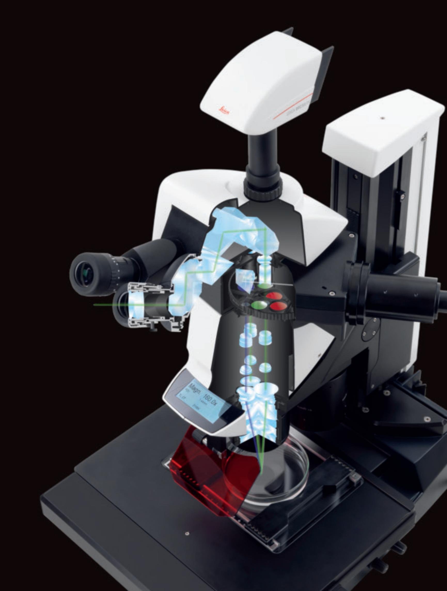
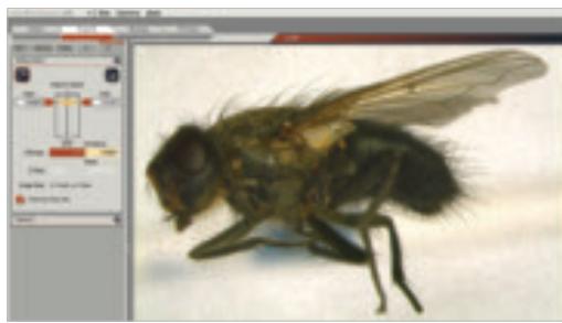
Until now, high depth of field and maximum resolution were always considered to be irreconcilable opposites. With FusionOptics™, Leica Microsystems has succeeded in overcoming these limitations. Scientific studies conducted at the Institute of Neuroinformatics, a department of the ETH Zürich, confirm that the human vision system is capable of drawing the maximum information content from each eye individually and merging it to create a three-dimensional image. In the same way, the new Leica M205 FA uses the two beam paths for different tasks: the right channel delivers a high-resolution image at the largest possible numerical aperture, while the left channel presents an image with high depth of field. As a result, two apparently irreconcilable worlds are merged in the human brain: the observer receives an image with outstanding richness of detail and outstanding depth of field at the same time.
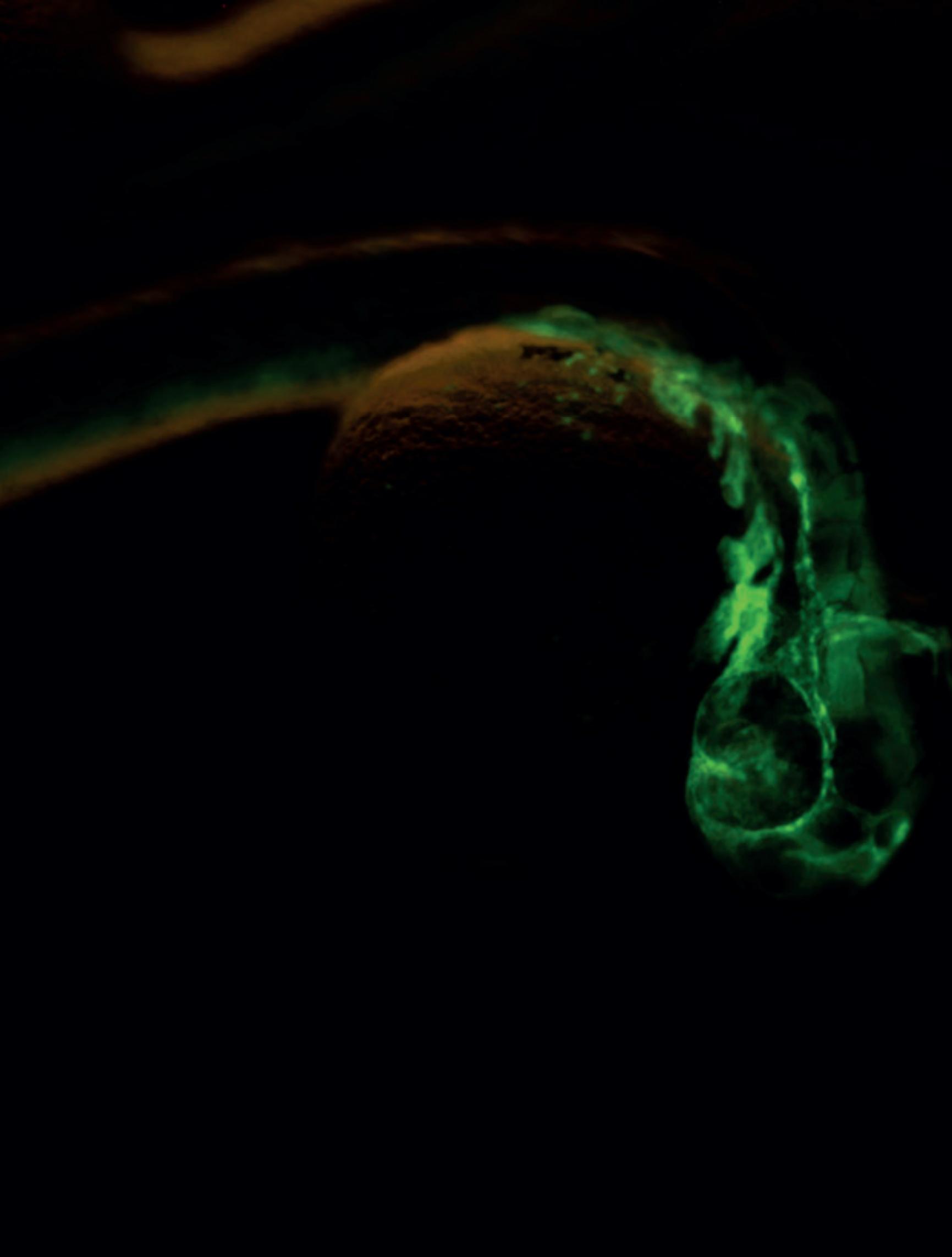
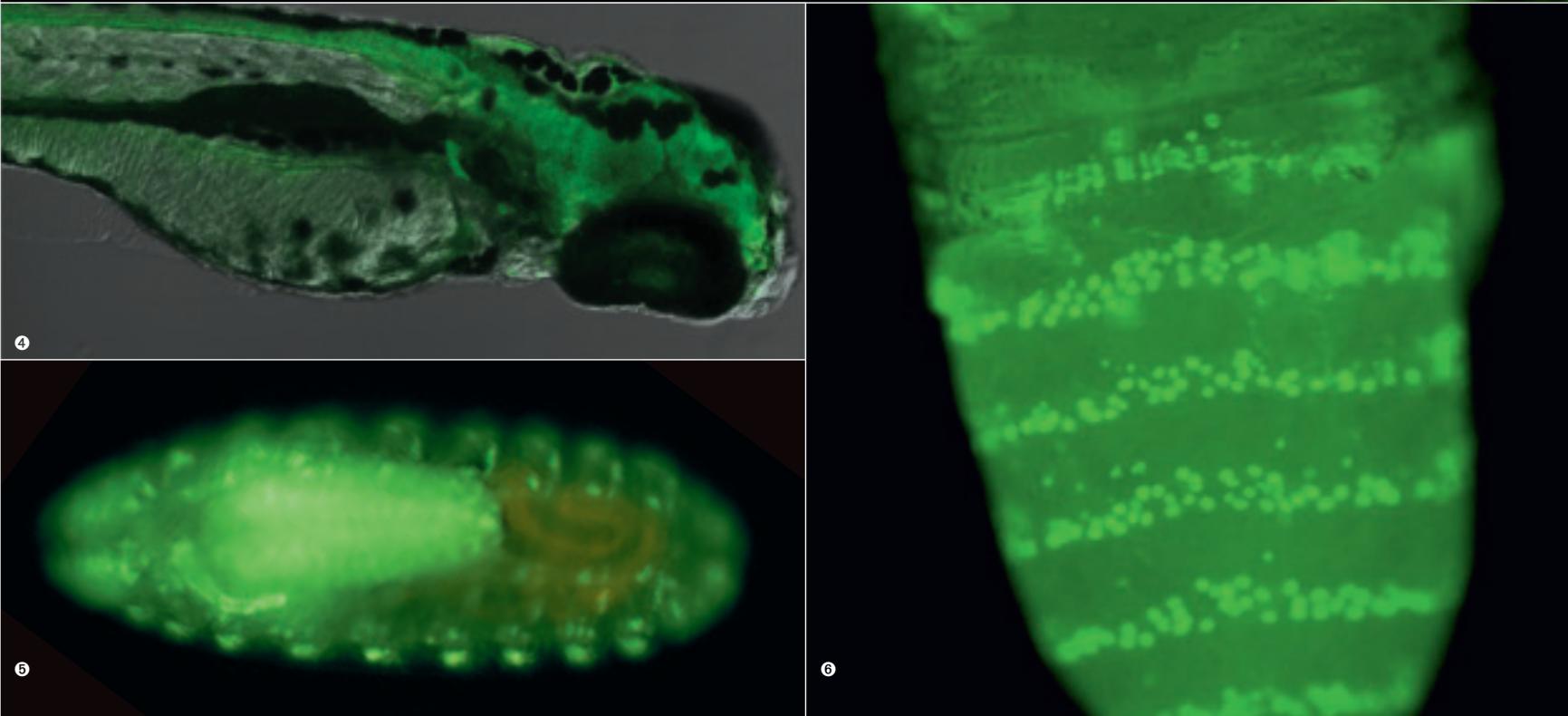
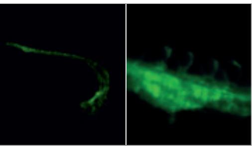
123 Vascular anatomy of a Zebrafish embryo as revealed by GFP expression driven by the Fli-1 promoter. Courtesy: Brant Weinstein, National Institutes of Health, Bethesda, MD
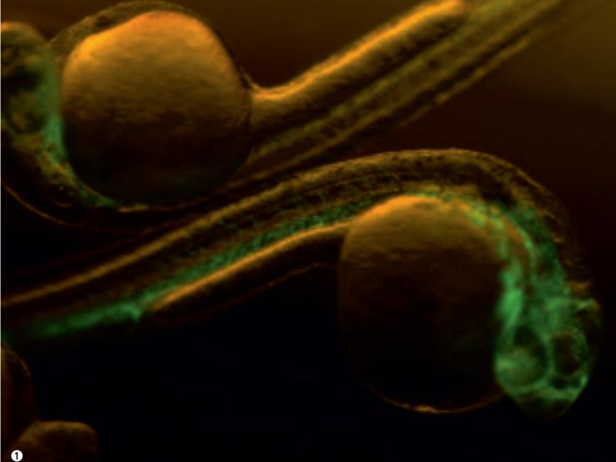
Zebrafish embryo expressing GFP under the control of the beta-actin promoter. Courtesy: Prof. Dr. Stephan C. F. Neuhauss, Professor for Neurosciences ETH Zurich and Institute for Brain Research at the University of Zurich
Periferic and central nervous (ventral cord) system of a drosophila embryo, salivary gland
Drosophila melanogaster. Dorsal view, Pupa; Green: Venus. Transgenic fluorescent protein in posterior compartment of each segment. Courtesy of Dr. Kuranaga, Dept. Genetics, Graduate School of Pharmaceutical Sciences, The University of Tokyo
The Art of Creating Brilliant Images
Illuminate specimens with patented third beam path
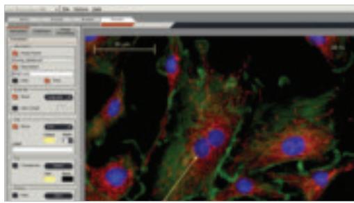
The Leica M205 FA and M165 FC stereomicroscopes feature Leica Microsystems' patented TripleBeam® technology. The TripleBeam® principle refers to the microscope's third beam path, reserved exclusively for fluorescence illumination to deliver evenly illuminated, reflex-free fields of view at all zoom settings. This separation of illumination and observation beam paths ensures brilliant fluorescence images, rich in detail and contrast, with the best light efficiency. Even weak fluorescence signals are displayed with remarkable image quality.
Optional Leica FluoCombi III™ for brilliant macro and micro imaging on one microscope
The unique Leica FluoCombi III™ attachment gives scientists the advantages of both stereo and high-resolution micro imaging ... on one microscope. By flipping a switch to activate the objective revolver, the user can quickly switch between a macro and a micro view of a specimen at any time.
In stereo mode, the system's large object field, working distance, and depth of field makes specimen manipulation easy. When the user is finished working in macro mode, he or she simply rotates the parcentric, parfocal micro objective into position. Viewing
performance up to 1500 line pairs per mm is achieved while retaining the exact focusing position. At the same time, the user can capture parallax-free z-stacks with maximum optical precision to obtain highly detailed 3-D information about the specimen.
Viewing the smallest detail to the entire organism, always in focus: the Leica FluoCombi III™ makes it easy to present research results with brilliant images.
Leica TripleBeam®: separate, third illumination path
Brilliant fluorescence
The best light efficiency
Leica FluoCombi III™: micro and macro views with one microscope
» Parallax-free documentation of the whole organism down to the smallest detail
Precisely detailed 3-D information
Microscopes that grow to meet future requirements
Adaptable to future experiments through maximum modularity
Seamless interaction of all system components
Leica M165 FC: Stereomicroscopy of the highest order
With Leica Microsystems' TripleBeam® technology, the Leica M165 FC Fluorescence Classic stereomicroscope documents the results of research with brilliant, contrast-rich, fluorescence images. The 16.5:1 zoom optics are fully apochromatically-corrected to resolve structures down to 551nm : classic, manual, high-level stereomicroscopy.
With an encoded zoom, filter changer, iris diaphragm, and objective revolver, the microscope configuration and optical data is available on a computer at any time. Experiment procedures and parameters are reproducible and consistent.
| Stereomicroscopy |
| FusionOptics™ |
| Zoom |
| Zoom range |
| Maximum magnification* |
| Max. objective aperture** |
| Max. resolution ** |
| Object field *** |
| Working distance *** |
| TripleBeam® path |
| Encoding **** |
| Complete automation |
| Four parfocal objectives |
| Objective nosepiece |
| FluoCombi III™ capable |
- With eyepieces 40 × and planapochromatic objective 2 ×
Planapo objective 2 ×
* Data with standard optics (objective 1×/ eyepieces 10×
**** Readout of settings for iris diaphragm, magnification, filter and objective in use per LAS (Leica Application Software)
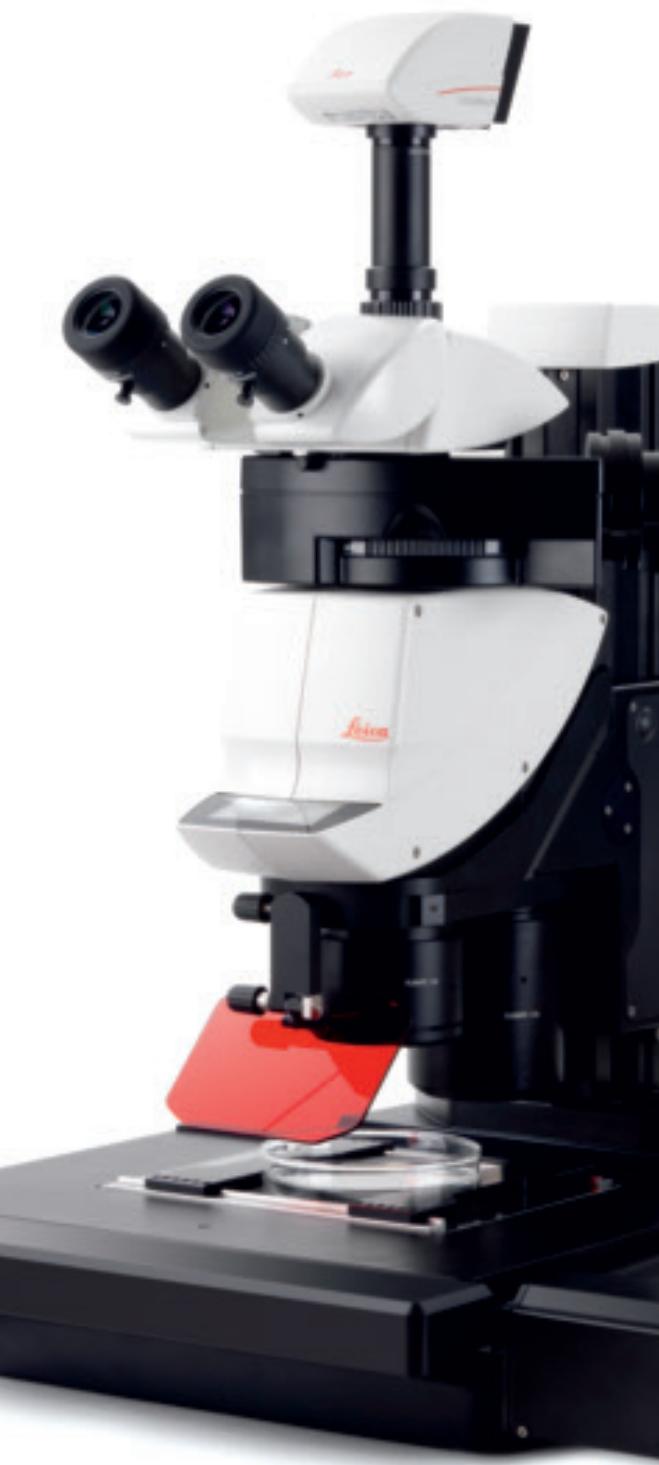
Leica M205 FA: Discover a New World of Research
| M165FC | M205FA |
| Manual | Complete Automation |
| No | Yes |
| 16.5:1 | 20.5:1 |
| 7.3×-120× | 7.8×-160× |
| 1920× | 2560× |
| 0.151 | 0.175 |
| 906 line-pairs/mm | 1050 line pairs/mm |
| 63 mm | 59 mm |
| 61.5 mm | 61.5 mm |
| Yes | Yes |
| Yes | Yes |
| No | Yes |
| Yes | Yes |
| Yes | Yes |
| Yes | Yes |
Leica Microsystems' unique combination of FusionOptics™ technology, TripleBeam® fluorescence, and an unprecedented level of microscope automation opens a new world of research with fluorescence stereomicroscopy. The fully apochromatically-corrected optics system; the largest zoom range available on the market, 20.5:1; and a resolution to 1050 line pairs per mm reveals a level of microscopic detail previously unknown in stereomicroscopy.
Time-intensive studies of live organisms and the resulting documentation of complex image sequences and multiple fluorescence images are easy to execute and are instantly reproducible by motorizing the focus, zoom, filter changer and iris diaphragm.
Concentrate the Experiment
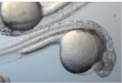
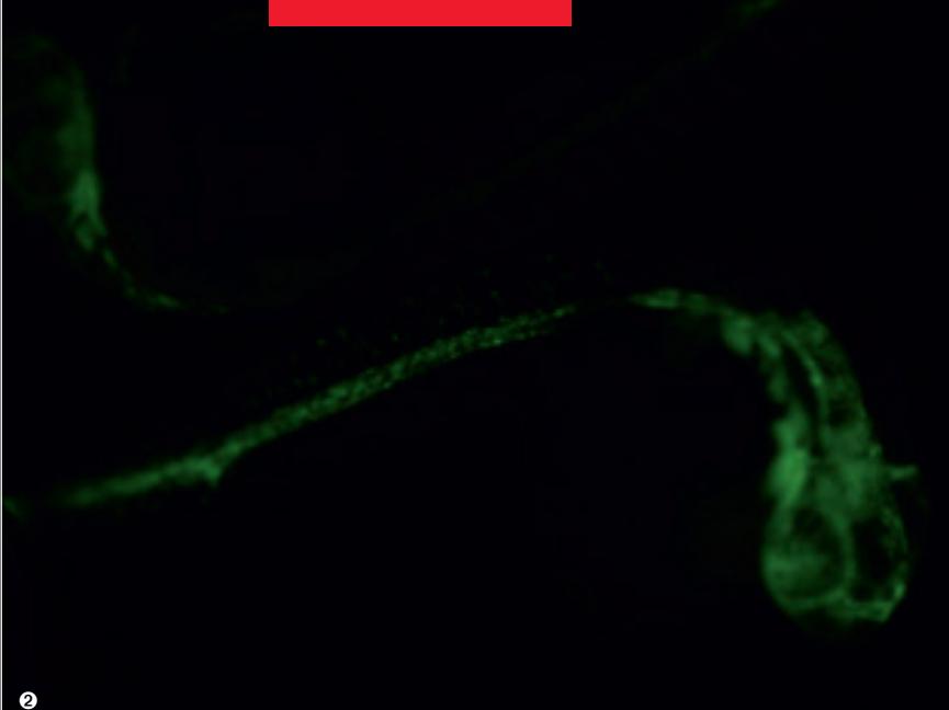
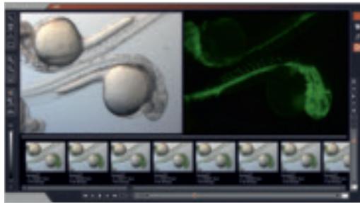
Zebrafish larva fin
The Leica IsoPro™ motorized cross-stage makes automated specimen scans easy.

With the touchscreen display, all important information and functions are at your fingertips.
Intelligent control
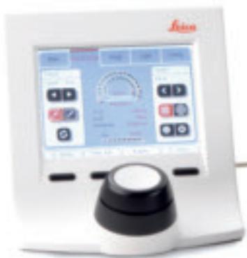
The user is in complete control of the experiment with just a few touches on the Leica SmartTouch™ control unit. The convenient, color touchscreen allows the user to save and recall all important optical parameters with a simple touch command on the visual controller display. The most important functions on the control unit can be customized to an individual's specific needs with freely programmable rotary knobs and memory function buttons. The design of the SmartTouch™ navigation display was designed to be ergonomic, intuitive, and efficient to limit the attention required to control the microscope and allow the user to focus entirely on the research and results.
Advanced life science applications
The researcher can control the Leica IsoPro™ motorized X/Y-stage dedicated for stereomicroscopy with the SmartTouch™ control unit, LAS software, or Leica AF6000 (Advanced Fluorescence) software. The user can easily reposition the microscope stage and program repetitive processes. The Leica M-Series Stereomicroscopes can expand into complete documentation systems for every requirement, from simple fluorescence photographs to intricate, multi-dimensional fluorescence experiments.
Investment for the future
Particularly in multi-user environments, an adaptable microscope system is important for meeting a wide range research requirements. The modular Leica M-Series platform features components and accessories work seamlessly together. The researcher can configure a tailormade stereomicroscope system for almost any research project, and have confidence that existing Leica Microsystems systems will adapt to scientific advances of the future.
The basis for successful documentation
Leica Microsystems offers a selection of powerful transmitted light bases that always present specimens in the best light: brightfield illumination with high or low diffusion, oblique transmitted light, and darkfield. The Rottermann Relief contrast method also ensures an excellent display, even when viewing unstained live cells.
Flexible Solutions for all Research Needs
Create the best spectral properties
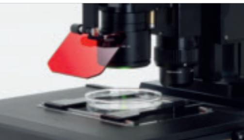
Leica Microsystems offers a wide range of microscope fluorescence filters that can be used with existing fluorescence filters to create the best spectral properties of a specimen. The Leica M-Series filter changer can accommodate up to four filter combinations (excitation and blocking filters). The fluorescence shutter does not open until a filter has been identified by its transponder in the observation channel. The shutter can be closed at any time with the press of a button to prevent over exposure of fluorescent light on to the specimens. In a software-controlled imaging series, the shutter only remains open during the image capture mode. This abbreviated shutter time and a filter change time of less than 500 ms are especially valuable for speeding up intensive fluorescence experiments.
Protect live cells
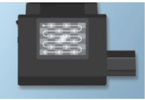
Protecting live cells and ensuring precise, constant culture conditions requires that an organism remains carefully controlled for an experiment's duration. The Leica MATS (Microscope stage Automatic Thermo control System) thermoplate radiates heat uniformly over the entire stage surface and precisely maintains a preset temperature. Constant temperature control helps ensure a successful experimental outcome.
Let there be light
The Leica EL6000 External Light source is equipped with a long-life metal halide lamp, a cost-effective, and timesaving alternative to mercury vapor lamps. Since this lamp does not need adjustment, the user is assured of uniformly illuminated, contrast-rich fluorescence images.
Ergonomically-designed stereomicroscope system
Leica Microsystems offers an unsurpassed range of observation tubes and ergomodules to configure the Leica M-Series. The new Leica Trinocular ErgoTube™ (5° to 45° observation angle) provides a wide range of adjustment options to provide a comfortable, relaxed seated position at the microscope. The Leica ErgoTube™ is designed to provide maximum comfort for all users, especially during long hours of work at the microscope.

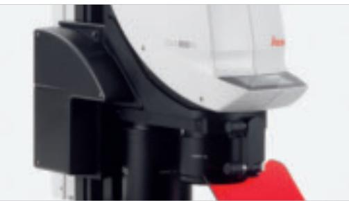
Assemble a fluorescence filter set to suite a specific application.
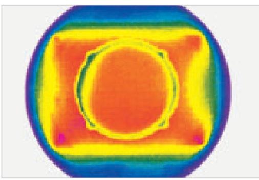
Leica MATS thermoplate: uniform temperature distribution for reliable experiment results.

The M-series stereomicroscope is adjustable even by millimeters to ensure comfortable work for hours.
Unprecedented Performance
Obtain overview and detailed image acquisition in 1 step.
Stability and ample space for all experimental situations.
Leica M165 FC fluorescence module.
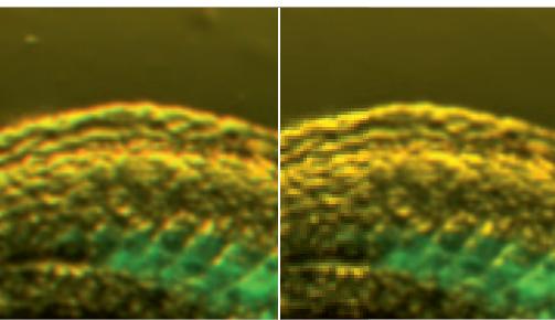
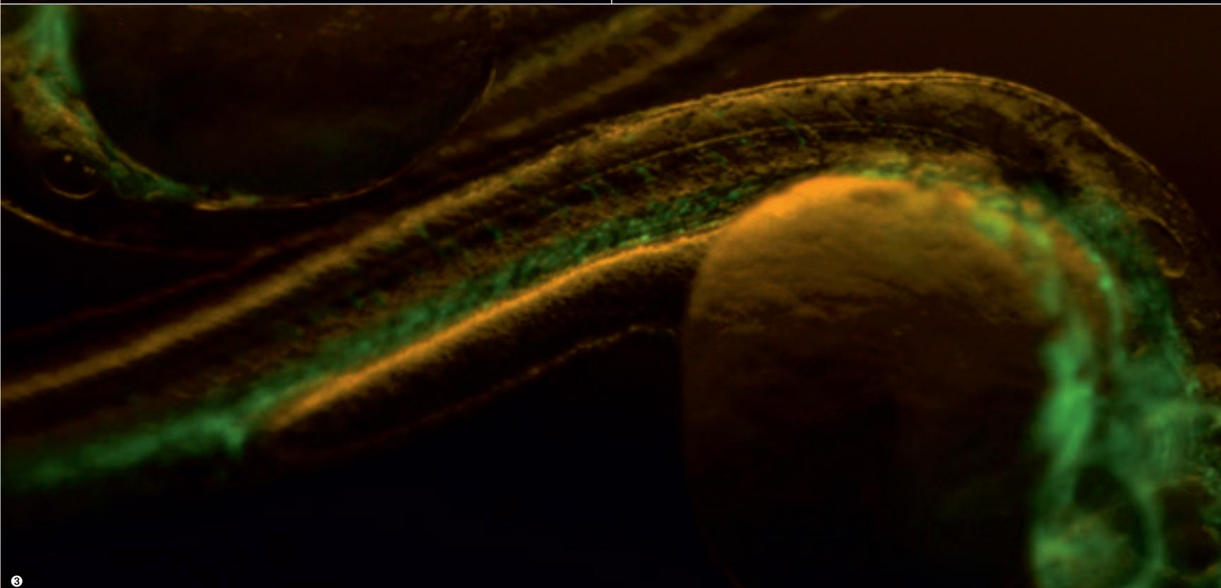
Uncorrected (left) and apochromatically-corrected (right) photo of a zebrafish larva.
Supreme performance for a broad range of research
The Leica M-Series Stereomicroscopes combine a large zoom range and high-resolution performance in a single system to enable a broad range of research tasks with just one microscope. For example, the researcher can not only observe organogenesis in an entire zebrafish but also cell diversification and determination in the retina.
The Leica M205 FA stereomicroscope advances research into magnification ranges previously unknown. With FusionOptics™ specimen details are clearly resolved down to a size of 476nm. The Leica M165 FC resolves structures down to a size of 551nm.
Space for your specimens
With the Leica M-Series high-performance stereomicroscopes, you no longer have to choose between a highly detailed presentation of a specimen and adequate space for manipulation. Four planapochromatically-corrected, parfocal main objectives can be used in any combination on the objective revolver. This gives enormous flexibility to choose the best magnification range and working distance to suit any application.
A solid basis for research
High-performance stereomicroscopes require a solid base. The M-Series construction is extremely stable to effectively absorb impact and vibration to virtually eliminate impaired image quality, even when observing specimens in a liquid medium.
Highly precise for complex imaging
Adjust the focal position of the microscope conveniently and precisely, even in the nanometer range, with the manual coarse/fine focus drive. Z-stacks and other complex multi-channel fluorescence imaging procedures are easy with the automated Leica M205 FA with motorized focus.
Enjoy the full performance capabilities
To provide all of the full performance capabilities of the new M-Series Stereomicroscopes, all system components are apochromatically-corrected. Fluorescence results are not marred by color seams or distortions. The new Leica M-Series reflects the superior imaging performance of Leica Microsystems' optical systems.
Fully-developed, Individually-tailored Solutions
The control center for experiments
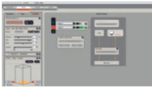
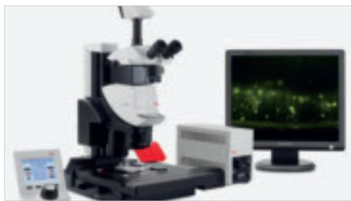
An automated, software-integrated M-Series system provides unprecedented convenience and simplifies experimental procedures, even in complex fluorescence applications. From controlling the microscope functions to capturing and processing images to analyzing and managing data: with Leica Microsystems, the microscope, camera, and software work in perfect harmony.
An integrated, complete imaging solution
Within the LAS environment, an automated stereomicroscope, digital camera, and software combine to create one user-friendly, consistent imaging solution. The versatility and modular design of LAS gives enormous flexibility to build an imaging system perfectly adapted to a unique application. LAS is an intuitive solution that makes both routine and research analysis easier.
Advanced software for fluorescence applications
Leica Microsystems has worked with leading scientists to develop Leica AF6000 Advanced Fluorescence software to meet every possible need in advanced life science applications. The intuitive software guides the user reliably and easily to brilliant results. For routine documentation, image superimposition, and time sequence imaging, Leica Microsystems offers the Leica AF6000 E introductory software package for fluorescence applications.
Leica AF6000 software fulfills all the requirements of fluorescence applications from multi-channel fluorescence to time sequence imaging, from z-stacks with parallax correction to 3-D image reconstruction. A motorized stage enables documentation of images on several selected areas of interest in the specimen. With many functions for documenting, quantifying, optimizing, and analyzing images, Leica AF6000 converts a stereomicroscope into an integrated, high-performance system that grows with your research needs.
LAS Multi-focus module
LAS Image Overlay module
AF6000: Settings for complex t and z series
AF6000: Image gallery in acquisition mode
Highlights of the
Leica M165 FC & M205 FA
Leica FusionOptics™ for never-before achieved depth of field and image brilliance.
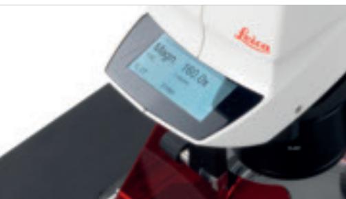
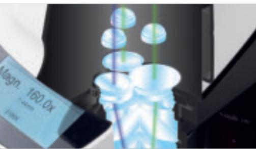
All data at a glance on the Leica M205 FA's display.
Easily switch between overview and parallax-free detailed view.

The new trinocular Leica ErgoTube™ quickly adapts to different users and various setups.
Fast and precise with reproducible zoom adjustment via the new motorized focus.
Never before seen: 3-D images of the highest resolution, brilliance, and depth of field
- FusionOptics™, integrated on the M205FA, with one channel for high resolution and one channel for high depth of field,
- create an image impression of unprecedented richness of detail and outstanding depth of field.
Largest zoom range on the market today
- Leica M205 FA: 20.5:1 zoom makes a wide range of research techniques possible with just one microscope.
Brilliant fluorescence images, rich detail and contrast
Patented Leica TripleBeam technology.
- Optimization of UV transmission of the illumination path.
- Filter properties adapt precisely to a specific specimen.
- Leica FluoCombi III™: Easy specimen manipulation with large working distance, depth impression, and field of view in stereo mode; capture parallax-free z-stacks with the micro objective for highly detailed, 3-D information.
Microscopes that grow to meet future requirements
- The Leica M-Series is a highly modular platform.
- A wide selection of accessories provides maximum system flexibility.
- The user can create individual filter combinations.
- The new Leica M-Series seamlessly integrates with existing system components.
- Trinocular Leica ErgoTube™: the best viewing comfort for different microscope users.
User comfort and experiment reproducibility through motorization
- Leica M205 FA: Easy documentation of complex image sequences and multiple fluorescence photos with motorized focus, zoom, filter changer, and iris diaphragm.
- Motorized Leica IsoPro™ cross-stage is ideal for complex, multi-dimensional fluorescence experiments.
Intelligent control with Leica SmartTouch™
- External control unit features clearly organized, colored touchscreen.
- Continuous status monitoring and convenient control of all system settings and functions.
- Individually program the most important control functions.
- Intuitive operation in seven different languages.
Encoding for experimental reproducibility and consistency
- Leica M165 FC features encoding of the focus, zoom, filters, and iris diaphragm.
- Microscope configuration and optical data and can be read out at a computer at any time.
Detailed view with high-performance objectives
- High-resolution, precisely-detailed view combines with a large working distance and ample space for specimen manipulation.
- Features four planapochromatically-corrected, parfocal main objectives.
- Convenient objective revolver provides a fast enlargement of the field of view
A solid foundation for research: stable construction
- The stable mechanical construction combines with superior optical performance for years of high-performance.
- Reliably absorbs impacts and vibrations to ensure outstanding image quality, even when observing specimens in a liquid medium.
Integrated solutions make research easy
- A seamless interface of components create a microscope system tailored to an individual application.
- From microscope control to image capture and processing to data analysis and management: the Leica M-Series provides flexibility, user friendliness, and reliable documentation of research results.
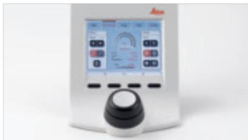
With Leica SmartTouch™, all motorized functions are at the fingertips, just a few clicks away.
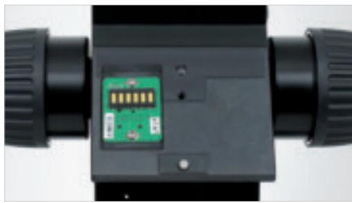
Contacts of internal instrument encoding.
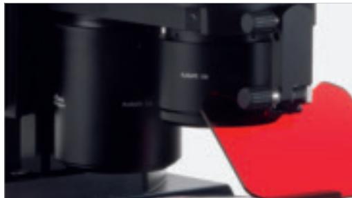
Fast objective change with the new objective turret.
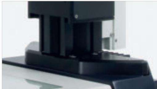
Stable mechanical construction supports superior optical performance.
Integrated, complete imaging solutions, by Leica Microsystems.
"With the user, for the user"
Leica Microsystems
Leica Microsystems operates globally in four divisions, where we rank with the market leaders.
- Life Science Division
The Leica Microsystems Life Science Division supports the imaging needs of the scientific community with advanced innovation and technical expertise for the visualization, measurement, and analysis of microstructures. Our strong focus on understanding scientific applications puts Leica Microsystems' customers at the leading edge of science.
- Industry Division
The Leica Microsystems Industry Division's focus is to support customers' pursuit of the highest quality end result. Leica Microsystems provide the best and most innovative imaging systems to see, measure, and analyze the microstructures in routine and research industrial applications, materials science, quality control, forensic science investigation, and educational applications.
- Biosystems Division
The Leica Microsystems Biosystems Division brings histopathology labs and researchers the highest-quality, most comprehensive product range. From patient to pathologist, the range includes the ideal product for each histology step and high-productivity workflow solutions for the entire lab. With complete histology systems featuring innovative automation and Novocastra™ reagents, Leica Microsystems creates better patient care through rapid turnaround, diagnostic confidence, and close customer collaboration.
Surgical Division
The Leica Microsystems Surgical Division's focus is to partner with and support surgeons and their care of patients with the highest-quality, most innovative surgical microscope technology today and into the future.
The statement by Ernst Leitz in 1907, "with the user, for the user," describes the fruitful collaboration with end users and driving force of innovation at Leica Microsystems. We have developed five brand values to live up to this tradition: Pioneering, High-end Quality, Team Spirit, Dedication to Science, and Continuous Improvement. For us, living up to these values means: Living up to Life.
Active worldwide
| Australia: | North Ryde | Tel. +61 2 8870 3500 | Fax +61 2 9878 1055 |
| Austria: | Vienna | Tel. +43 1 486 80 50 0 | Fax +43 1 486 80 50 30 |
| Belgium: | Groot Bijngaarden | Tel. +32 2 790 98 50 | Fax +32 2 790 98 68 |
| Canada: | Richmond Hill/Ontario | Tel. +1 905 762 2000 | Fax +1 905 762 8937 |
| Denmark: | Herlev | Tel. +45 4454 0101 | Fax +45 4454 0111 |
| France: | Nanterre Cedex | Tel. +33 811 000 664 | Fax +33 1 56 05 23 23 |
| Germany: | Wetzlar | Tel. +49 64 41 29 40 00 | Fax +49 64 41 29 41 55 |
| Italy: | Milan | Tel. +39 02 574 861 | Fax +39 02 574 03392 |
| Japan: | Tokyo | Tel. +81 3 5421 2800 | Fax +81 3 5421 2896 |
| Korea: | Seoul | Tel. +82 2 514 65 43 | Fax +82 2 514 65 48 |
| Netherlands: | Rijswijk | Tel. +31 70 4132 100 | Fax +31 70 4132 109 |
| People's Rep. of China: | Hong Kong | Tel. +852 2564 6699 | Fax +852 2564 4163 |
| Portugal: | Lisbon | Tel. +351 21 388 9112 | Fax +351 21 385 4668 |
| Singapore | Tel. +65 6779 7823 | Fax +65 6773 0628 | |
| Spain: | Barcelona | Tel. +34 93 494 95 30 | Fax +34 93 494 95 32 |
| Sweden: | Kista | Tel. +46 8 625 45 45 | Fax +46 8 625 45 10 |
| Switzerland: | Heerbrugg | Tel. +41 71 726 34 34 | Fax +41 71 726 34 44 |
| United Kingdom: | Milton Keynes | Tel. +44 1908 246 246 | Fax +44 1908 609 992 |
| USA: | Bannockburn/llinois | Tel. +1 847 405 0123 | Fax +1 847 405 0164 |
and representatives in more than 100 countries
In accordance with the ISO 9001 certificate, Leica Microsystems (Switzerland) Ltd, Industry Division, has at its disposal a management system that meets the requirements of the international standard for quality management. In addition, production meets the requirements of the international standard ISO 14001 for environmental management.
- Bringing Ideas into the Light
- FusionOptics™:
- The Evolution of Resolution
- FusionOptics
- Leica Microsystems brings high resolution and depth of field together
- The Art of Creating Brilliant Images
- Illuminate specimens with patented third beam path
- Optional Leica FluoCombi III™ for brilliant macro and micro imaging on one microscope
- Leica TripleBeam®: separate, third illumination path
- Leica FluoCombi III™: micro and macro views with one microscope
- Microscopes that grow to meet future requirements
- Leica M165 FC: Stereomicroscopy of the highest order
- Leica M205 FA: Discover a New World of Research
- Concentrate the Experiment
- Intelligent control
- Advanced life science applications
- Investment for the future
- The basis for successful documentation
- Flexible Solutions for all Research Needs
- Create the best spectral properties
- Protect live cells
- Let there be light
- Ergonomically-designed stereomicroscope system
- Unprecedented Performance
- Supreme performance for a broad range of research
- Space for your specimens
- A solid basis for research
- Highly precise for complex imaging
- Enjoy the full performance capabilities
- Fully-developed, Individually-tailored Solutions
- The control center for experiments
- An integrated, complete imaging solution
- Advanced software for fluorescence applications
- Highlights of the
- Leica M165 FC & M205 FA
- Never before seen: 3-D images of the highest resolution, brilliance, and depth of field
- Largest zoom range on the market today
- Brilliant fluorescence images, rich detail and contrast
- User comfort and experiment reproducibility through motorization
- Intelligent control with Leica SmartTouch™
- Encoding for experimental reproducibility and consistency
- Detailed view with high-performance objectives
- A solid foundation for research: stable construction
- Integrated solutions make research easy
- "With the user, for the user"
- Leica Microsystems
- - Life Science Division
- - Industry Division
- - Biosystems Division
- Surgical Division
- and representatives in more than 100 countries
Brand : LEICA
Model : M205 FA
Category : Microscope