
M205 A - Microscope LEICA - Notice d'utilisation et mode d'emploi gratuit
Retrouvez gratuitement la notice de l'appareil M205 A LEICA au format PDF.

| Type de produit | Microscope stéréoscopique motorisé haut de gamme |
| Zoom | 20,5:1 (premier au monde) |
| Résolution maximale | 1050 paires de lignes/mm (structures < 476 nm) |
| Ouverture numérique | 0,35 (avec objectif 2× planapochromatique) |
| Distance de travail | 61,5 mm (avec objectif 1×) |
| Optiques | Correction apochromatique, FusionOptics™ (canal droit haute résolution, canal gauche grande profondeur de champ) |
| Motorisation | Zoom, mise au point et diaphragme iris motorisés, commande numérique |
| Éclairage | Compatible LED5000 MCI™ (contraste multi) et LED5000 RL (anneau), intégrés et contrôlables via LAS |
| Logiciel | Leica Application Suite (LAS) pour acquisition, analyse et documentation |
| Codage électronique | Lecture continue de la magnification, transmission à LAS |
| Oculaires | Tube trinoculaire, angle d'observation 5°–45° réglable |
| Correction dioptrique | Réglable avec système de verrouillage |
| Champ numérique | 23 mm (grand champ) |
| Colonne de mise au point | Extrêmement stable, qualité pour hauts grossissements |
| Modularité | Compatible avec une large gamme d'objectifs, statifs, caméras et accessoires |
| Entretien et nettoyage | Nettoyer les lentilles avec un chiffon doux non pelucheux ; éviter les solvants agressifs. Maintenir l'instrument dans un environnement sec et propre. |
| Sécurité | Utiliser uniquement avec des accessoires d'origine ; ne pas exposer à l'humidité ou à des températures extrêmes. Débrancher avant entretien. |
| Pièces détachées et réparabilité | Contacter Leica Microsystems ou un distributeur agréé pour toute réparation ou pièce de rechange. |
FOIRE AUX QUESTIONS - M205 A LEICA
Questions des utilisateurs sur M205 A LEICA
0 question sur cet appareil. Repondez a celles que vous connaissez ou posez la votre.
Poser une nouvelle question sur cet appareil
Téléchargez la notice de votre Microscope au format PDF gratuitement ! Retrouvez votre notice M205 A - LEICA et reprennez votre appareil électronique en main. Sur cette page sont publiés tous les documents nécessaires à l'utilisation de votre appareil M205 A de la marque LEICA.
MODE D'EMPLOI M205 A LEICA
Leica M205 A, M205 C,
M165 C & M125
Experience another dimension in stereomicroscopy with the new, high performance stereomicroscopes from Leica.
Living up to Life
A Step Towards Infinity
Ever since their introduction by Horatio S. Greenough, stereomicroscopes have worked according to the optical principles based primarily on Ernst Abbe's research. For over a century, ingenious optics designers and engineers have worked to push magnification, resolution and image fidelity to the limit permitted by optics. In doing so, they have always been constrained by the interrelation between three factors: the higher a microscope's resolution, the lower the available working distance. If one increases the distance of the optical axes, the three-dimensional image seen by the observer becomes distorted a cube then becomes a tower, a flat surface curves towards the observer.
Limits are made to be broken.
LeicaM205A and M205 C are the first stereomicroscopes in the world that can offer a 20.5:1 zoom. This accomplishment, however, was not enough for Leica's engineers. With the new FusionOptics™, they have succeeded in taking yet another step beyond previous limits. Besides the greater magnification, they have also increased the resolution to 1050lp/mm, which corresponds to a resolved structure size of 476nm.
Of course, this performance increase is reflected in your daily work: Set up your specimens on the microscope table with comfortable freedom of movement and discover details that were previously unrevealed, even in stereomicroscopy.
The human brain is a fascinating piece of work. Using the data it receives from both eyes, it calculates a three-dimensional image of our environment in an unceasing stream. What is truly remarkable, however, is the brain's ability to gauge situations with lightning speed based on the information it receives, and to react to such situations appropriately.
Brain jogging with the Leica M205 A and M205 C
The new Leica M205 A and M205 C stereomicroscopes with FusionOptics™ rely on the unsurpassed adaptability of the brain. The microscope assigns a different task to each of the two beam paths: the right channel contains an image with very high resolution, while the left channel provides very high depth of field. The brain then automatically gathers the best information from both sources and uses it to compose one image with very high richness of detail and depth of field.

Juggling Increases Brain Size
Previous studies assumed that humans build brain mass during childhood, develop neurological networking through training in adolescence, can at best maintain this complexity during midlife, and will unavoidably experience diminished mental performance with increasing age.
Now, a study led by Dr. Arne May* of the University of Regensburg has shown that certain regions of the adult brain have the ability to build brain matter through training. In a group of laypersons who practiced juggling over a three-month period, structural changes in the cerebral cortex were identified after the training period. Astonishingly, the new brain matter formed primarily in the two areas that are responsible for vision and touch. Obviously, the difficulty in juggling lies in visually capturing and analyzing the balls' movements.
Leica FusionOptics™ takes advantage of the flexibility of our brains, and as an added benefit improves mental performance capability.
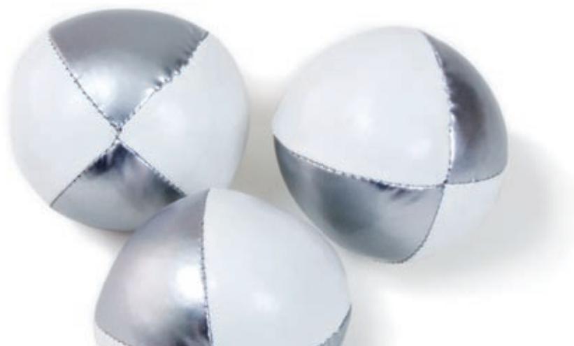
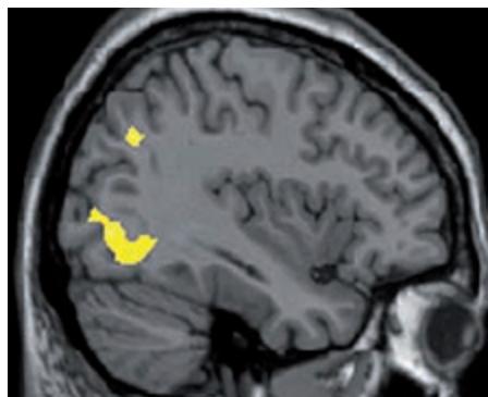

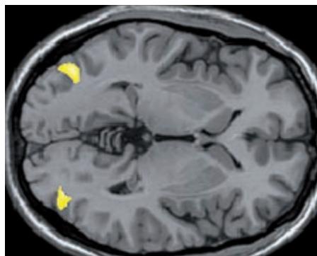
The areas marked in yellow are the regions in which new brain matter was shown to have been created. Courtesy of Dr. Arne May (University Clinic Hamburg)
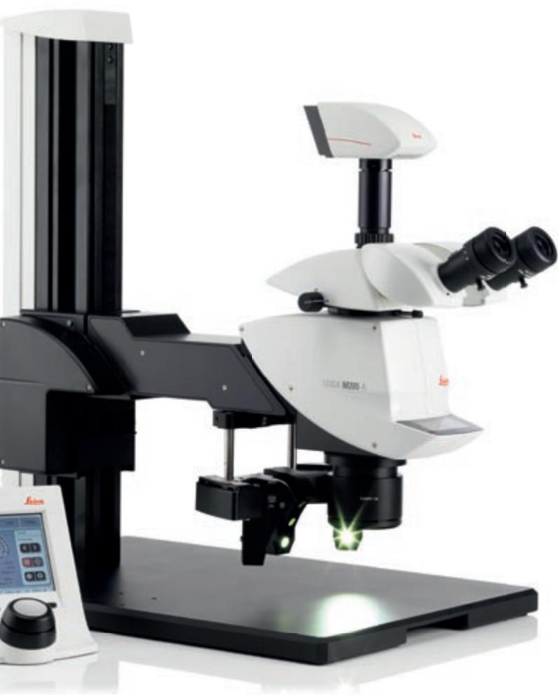
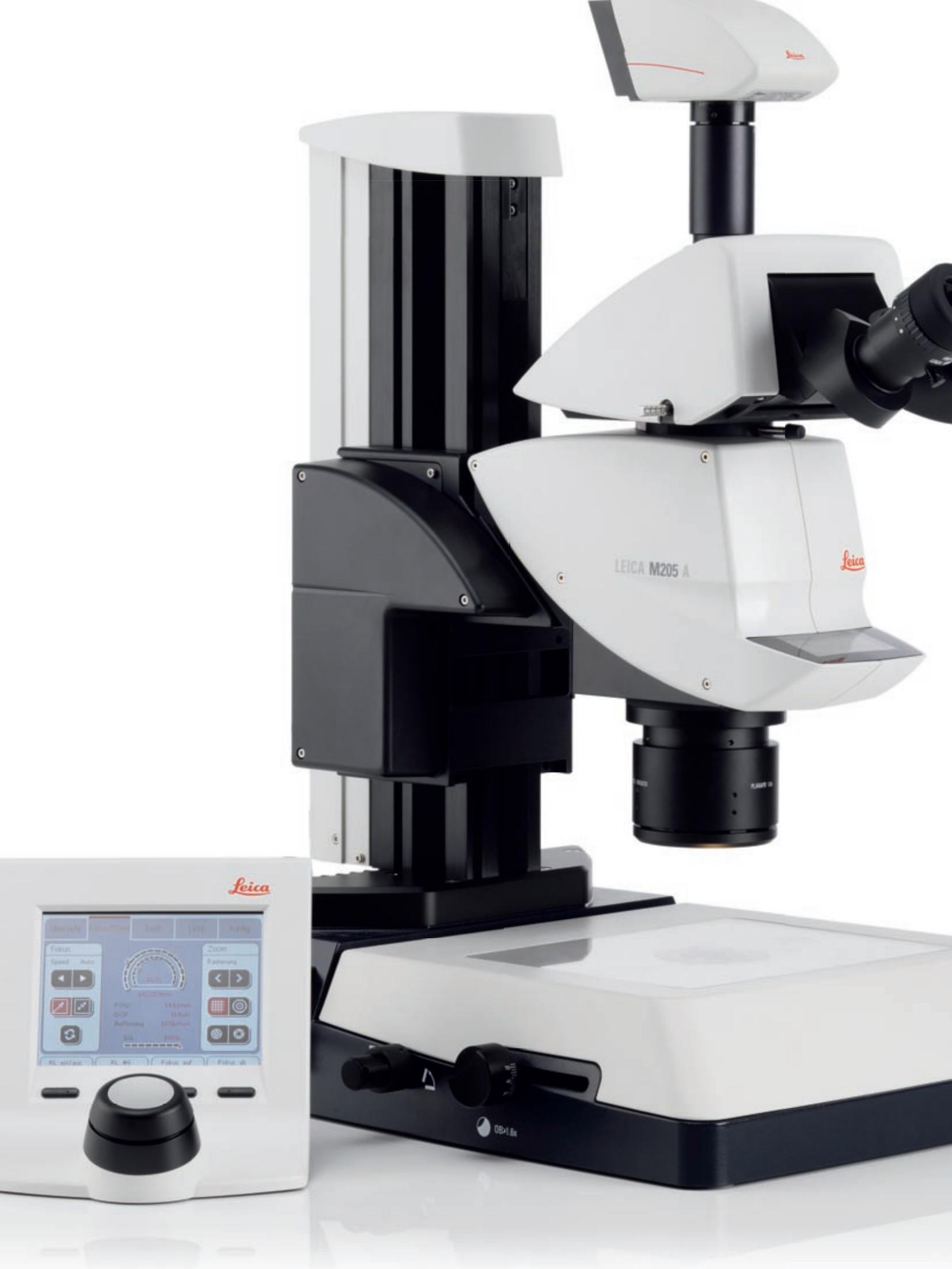
Leica M205 A: the top-of-the-line stereomicroscope for the fully automated complete system
With digitally controlled zoom, focus (with the new motorized focus drive) and iris diaphragm, a Leica DFC camera, the motorized mechanical stage and versatile software modules of the Leica Application Suite (LAS), the Leica M205 A is capable of accepting any settings and performing any analysis expected of a stereomicroscope with a few clicks of the mouse.
Of course, this directly affects your daily work: frequently used microscope settings can be restored with a few mouse clicks, time-consuming serial examinations, of relatively large specimens, can be programmed once in the computer and can then be allowed to run automatically whenever necessary.
Leica M205 C: advance into unexplored territory with
FusionOpticsTM
It does not matter if you need a large work surface with lots of room for handling specimens or analyzing the tiniest details, which were previously detectable only with a light microscope: with the extraordinary FusionOptics™ zoom, Leica Microsystems has set a new standard for stereomicroscopy.
What was previously thought to be an optical impossibility is now reality in the Leica M205 C. The zoom range of 7.8 × -160 × , objectives from 0.63 × -5 × , and an enormous selection of accessories make this a system that performs superbly in any application.
The New Leica M-Series: A Solution
for Every Task
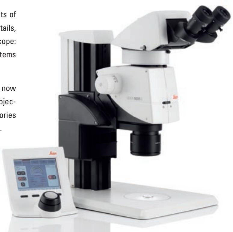
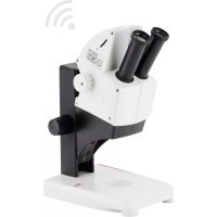
Leica M165 C: Classic stereomicroscopy of the highest order
For those who wish to continue working with classic stereomicroscopes, the Leica M165 C is equal to almost every need. The optical principle of two symmetrical channels is the same as that of the Leica MZ16, but the zoom range and numerical aperture have been increased to 16.5:1.
Of course, the Leica M165 C is also compatible with the full range of cameras, objectives, tubes, bases and accessories. This means not only that you are sure to find a configuration solution for almost any task you have now, but you can also be confident that you will always be able to take advantage of the latest advances in the Leica M series in the future.
Leica M125: one instrument for many tasks
Supreme performance is not always the most important requirement: in many routine situations, you simply need a microscope that is exceptionally tough and reliable, and compatible with a wide range of accessories. Of course one feature cannot be compromised: the best possible optical quality.
With a magnification range of 8 × -100 × , the Leica M125 is ideal for many applications that may be useful to you: from presorting mechanical components to analyzing plastics and detailed inspection of printed circuit boards, the Leica M125 provides consistently high-quality, detailed images of your specimens.
You Can Have It All at Once
High magnification with great ergonomic benefits
Conventional stereomicroscopy gives users a choice: they must choose high resolutions and richness of detail, or opt for a larger working distance to be able to manipulate the work specimen. The higher a microscope's resolution, the less free room there is between specimen and objective.
The Leica M205 A and M205 C uses one 1 × objective to advance into magnification ranges that were previously only possible with high magnification objectives. This has a directly positive influence on your daily work: the Leica M205 A and M205 C can resolve structures of less than one micrometer. At the same time, the user has a clearance of 61.5mm manipulating specimens. Sorting and processing even for the smallest details can be carried out easily without changing objectives.
APO for all
To take full advantage of the performance capabilities of these new instruments, all new M-series components are corrected exclusively apochromatically. At last, color fringes can be consigned to history once and for all.
Up to Your Tasks. Put Us to the Test!

Whenever human lives may be affected, technology must be 100% reliable. For this reason, autopilots in airplanes and trigger mechanisms in airbags, for example, are subjected to stringent materials testing. This testing includes a wide variety of methods used to measure the behavior and material characteristics of standardized material samples or finished parts (component testing) under thermal, mechanical and chemical stress. During the tests, a material is checked for purity, faults or load capacity.
With the Leica M205 A, M205 C, M165 C and M125 stereomicroscopes, experts in the materials test laboratory can delve into even smaller structures. Thanks to the unique FusionOptics™ system on the Leica M205 A and M205 C, for the first time ever in stereomicroscopy, increased depth of field can be combined with higher resolution.
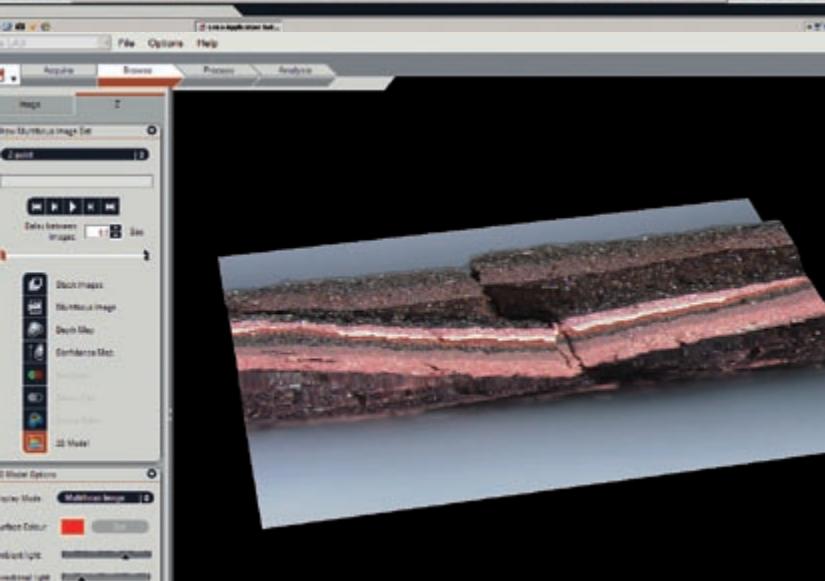
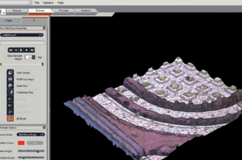
With the electronic coding that is applied for all components involved in imaging, the tester can eliminate practically all sources of error associated with measurement applications, and reproduce his test settings at any time. Using the Leica Application Suite (LAS), you can read out all relevant data, store images in an image database and measure and analyze them using the computer. The two new light sources, LED5000 MCI™ (Multi Contrast Illumination) and LED5000 RL offer a wide variety of adjustment and automation options. Along with the automated mechanical stage and the new M-series, they form a complete system. Not only does this help to avoid errors, it also saves time, one of the most important advantages offered by the new Leica M205 A, M205 C, M165 C and M125.
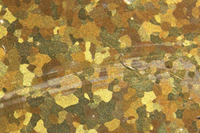
Brush marks on metal section
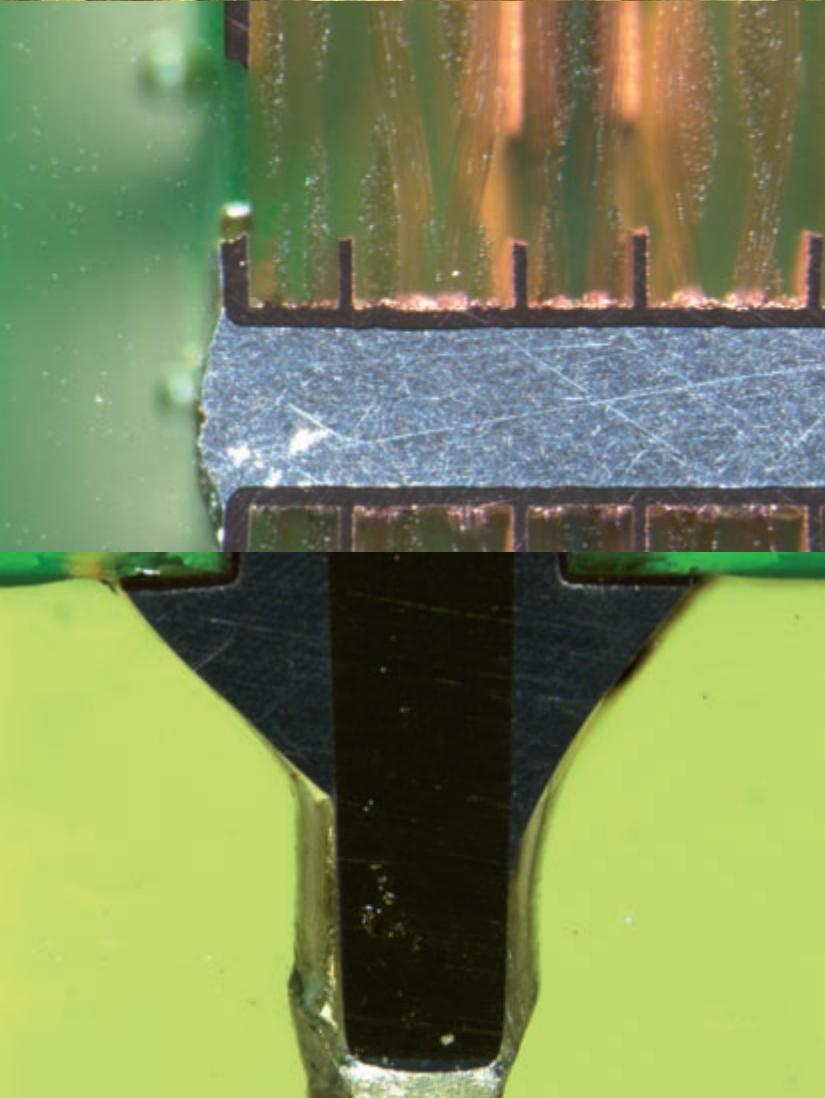
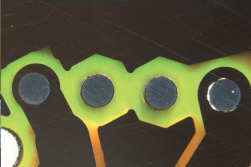
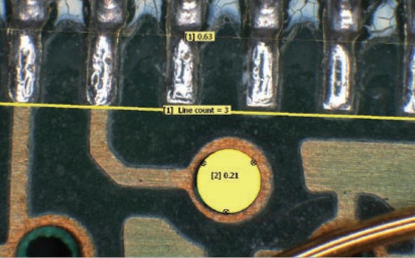
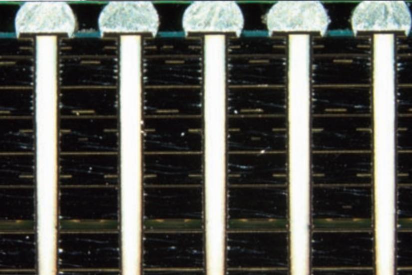
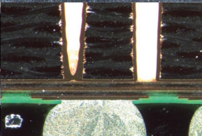
Through-connections on printed circuit board
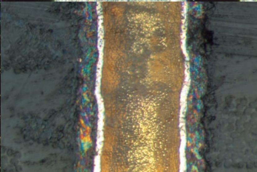
Section through soldered joint
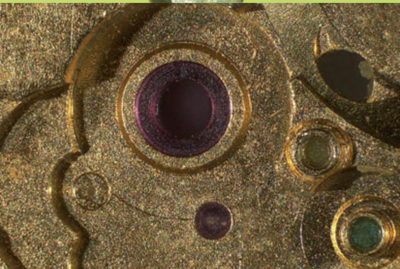
Cutout of clockwork
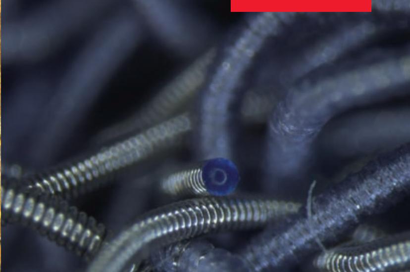
Medical implant
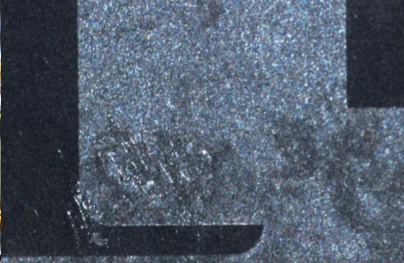
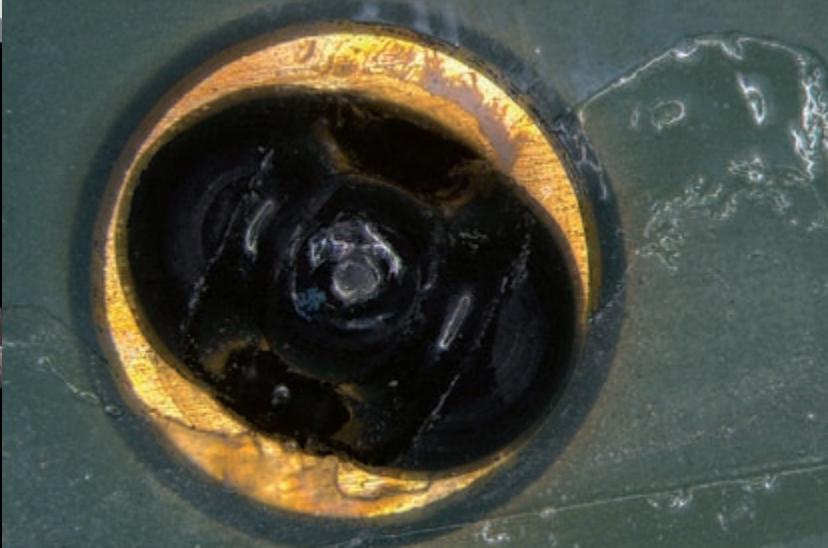
Defective through-connection
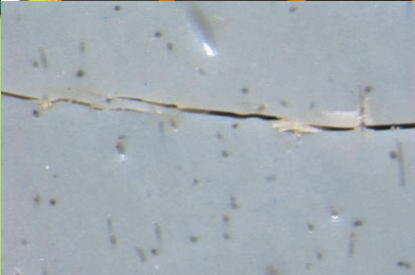
Microcrack through composite material
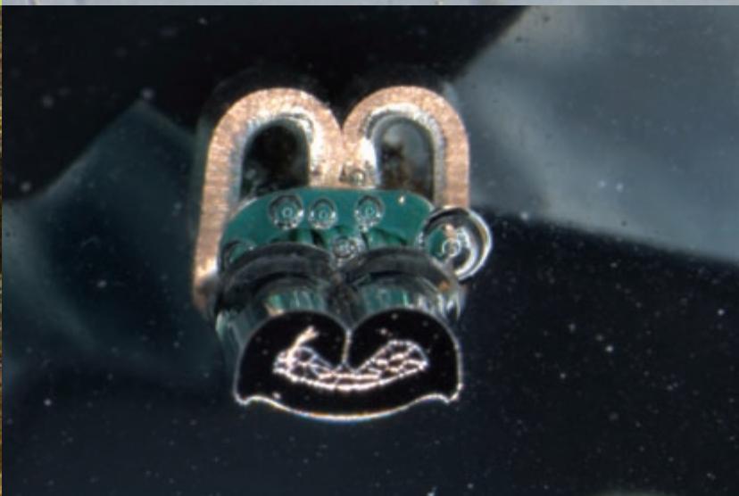
Cut through crimp contact
Leica Application Suite: the Cerebrum for Your Data
Integrated complete solution
The Leica Application Suite (LAS) combines automated microscopes, digital cameras, illumination and software from Leica in a single environment for a consistent, user-friendly imaging solution with incomparable performance. Thanks to its versatility, the Leica Application Suite can be used for an exceptionally wide variety of applications. With its wide range of image processing functions, the LAS cuts down the time required for displaying, processing, measuring and documenting digital images. The software monitors all Leica components that are connected to the computer, such as the stereomicroscope, objective changer, DFC camera, LED5000 illumination and motorized cross-stage. The data thus obtained are processed in LAS; to do so, all installed modules communicate with each other. Thus LAS is an intuitive solution that makes both routine and research analysis easier.
Features at a glance:
- LAS increases productivity by integrating microscopes, digital cameras and application software in order to coordinate imaging tasks using an intelligent control system.
LAS automates the digital microscopy environment with the computer-aided functions of Leica microscopes. - Just one application does everything: recording, storing and commenting; it can also display high-quality images in a thumbnail gallery.
- High modularity – software that keeps pace with your tasks.
Technical Highlights
Leica M205 A, M205 C, M165 C, and M125
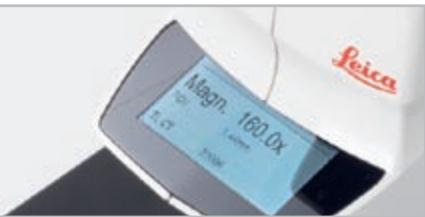
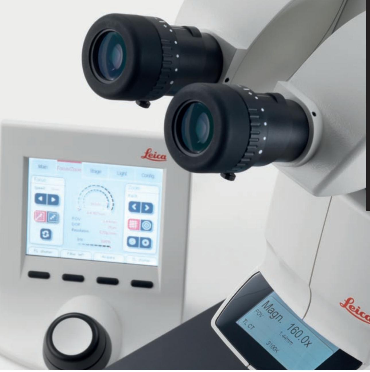
The digital display of the Leica M205 A shows allessential settings at a glance
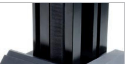
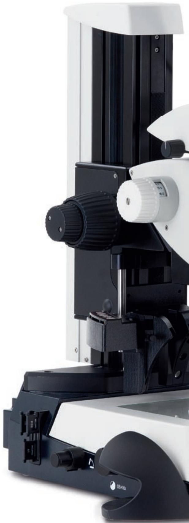
A new, extremely stable focusing column ensures the quality of high magnification
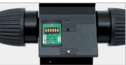
Contacts of internal instrument encoding
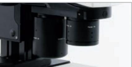
The objective nosepiece also conforms to the highest standards applied to the magnification range without tedious refocusing
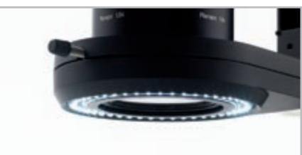
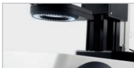
The LED5000-RL ring light is one of the new, fully integrated illumination components and can be controlled entirely on the device itself or via the Leica Application Suite
Stereomicroscope with the highest zoom
20.5:1 zoom allows overview and detail observation using one instrument
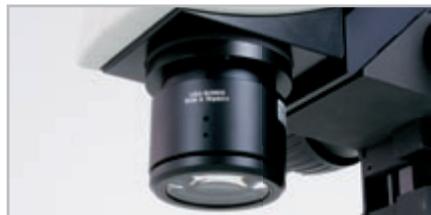
Numerical aperture of 0.35 (with 2 × planapochromatic objective)
One-of-a-kind resolution of 1050lp/mm enables resolution of structures smaller than 476nm
Rigid, sturdy mechanical structure
Rigid, sturdy structure supports high optical performance
- Detail solutions such as integrated cable duct and complete integration of electronics keep your workspace neat and clean
Encoding and Motorization
- M165 C / M205 C: Continuous electronic readout of the magnification
M205 A: Motorization of magnification and iris diaphragm
Parfocal objective nosepiece
Objective changes without refocusing
- User-defined combination of main objectives provides huge range of applications
- Encoding provides continuous configuration information to the LAS
Modularity
- New Leica M-series instruments can be combined with many existing system components
- Selection of various objectives, stands, cameras, illumination sources and other accessories
- The optimal solution is assured for practically any application
Completely integrated illuminator
- New illumination components seamlessly integrated in the complete system
- Complete control and reproducibility of settings
- Finding the right illumination setting is a snap
- Complete control of settings, manually or using LAS
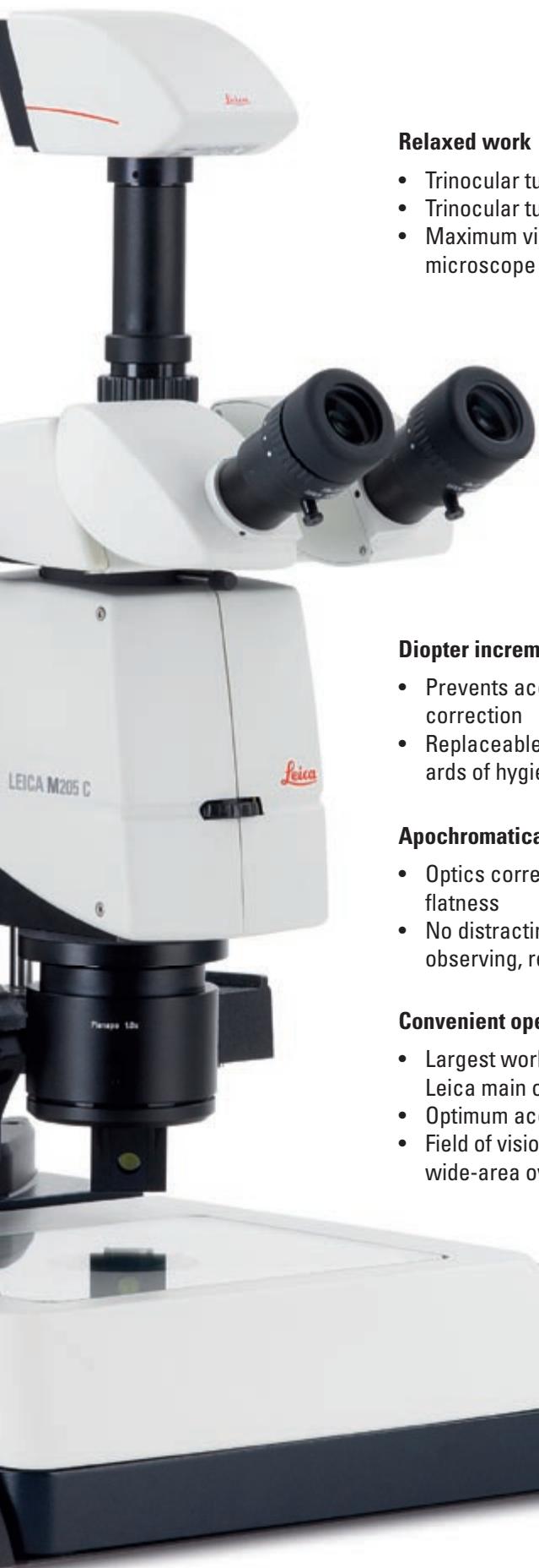
Relaxed work
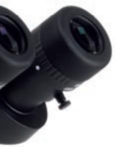
- Trinocular tube with 30^ viewing angle
Trinocular tube with 5^ - 45^ viewing angle
Maximum viewing comfort for different microscope users
Revolutionary FusionOptics™ (Leica M205 C)
Right channel with high resolution
- Left channel with high depth of field
Information from both channels is combined in the brain
Unparalleled resolution, brilliance and depth of field
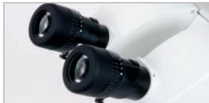
Diopter increments
Prevents accidental adjustment of dioptic correction
- Replaceable eyecups for the highest standards of hygiene
Apochromatically corrected optics
- Optics corrected for chromatic aberrations and flatness
- No distracting color fringes or distortions when observing, recording or analyzing images
Convenient operation under the microscope
- Largest working distances for all Leica main objectives
Optimum access to work specimen
Field of vision number 23 enables wide-area overview of the specimen
Leica M125 with new motorized focus and LED5000 RL LED illumination

The viewing angle is adjustable from 5^ - 45^ for the most relaxed head position possible
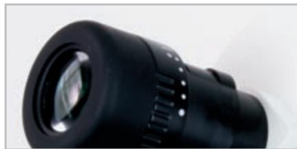
A locking system on the oculars prevents the diopter compensation from being changed inadvertently
The new, planapochromatic objectives prevent color seams, and field number 23 enables a large overview of the specimen
Planapo 1 × : the working distance of 61.5mm leaves a lot of free space under the objective
"With the user, for the user"
Leica Microsystems
Leica Microsystems operates globally in four divisions, where we rank with the market leaders.
- Life Science Division
The Leica Microsystems Life Science Division supports the imaging needs of the scientific community with advanced innovation and technical expertise for the visualization, measurement, and analysis of microstructures. Our strong focus on understanding scientific applications puts Leica Microsystems' customers at the leading edge of science.
- Industry Division
The Leica Microsystems Industry Division's focus is to support customers' pursuit of the highest quality end result. Leica Microsystems provide the best and most innovative imaging systems to see, measure, and analyze the microstructures in routine and research industrial applications, materials science, quality control, forensic science investigation, and educational applications.
- Biosystems Division
The Leica Microsystems Biosystems Division brings histopathology labs and researchers the highest-quality, most comprehensive product range. From patient to pathologist, the range includes the ideal product for each histology step and high-productivity workflow solutions for the entire lab. With complete histology systems featuring innovative automation and Novocastra™ reagents, Leica Microsystems creates better patient care through rapid turnaround, diagnostic confidence, and close customer collaboration.
Surgical Division
The Leica Microsystems Surgical Division's focus is to partner with and support surgeons and their care of patients with the highest-quality, most innovative surgical microscope technology today and into the future.
The statement by Ernst Leitz in 1907, "with the user, for the user," describes the fruitful collaboration with end users and driving force of innovation at Leica Microsystems. We have developed five brand values to live up to this tradition: Pioneering, High-end Quality, Team Spirit, Dedication to Science, and Continuous Improvement. For us, living up to these values means: Living up to Life.
Active worldwide
| Australia: | North Ryde | Tel. +61 2 8870 3500 | Fax +61 2 9878 1055 |
| Austria: | Vienna | Tel. +43 1 486 80 50 0 | Fax +43 1 486 80 50 30 |
| Belgium: | Groot Bijngaarden | Tel. +32 2 790 98 50 | Fax +32 2 790 98 68 |
| Canada: | Richmond Hill/Ontario | Tel. +1 905 762 2000 | Fax +1 905 762 8937 |
| Denmark: | Herlev | Tel. +45 4454 0101 | Fax +45 4454 0111 |
| France: | Nanterre Cedex | Tel. +33 811 000 664 | Fax +33 1 56 05 23 23 |
| Germany: | Wetzlar | Tel. +49 64 41 29 40 00 | Fax +49 64 41 29 41 55 |
| Italy: | Milan | Tel. +39 02 574 861 | Fax +39 02 574 03392 |
| Japan: | Tokyo | Tel. +81 3 5421 2800 | Fax +81 3 5421 2896 |
| Korea: | Seoul | Tel. +82 2 514 65 43 | Fax +82 2 514 65 48 |
| Netherlands: | Rijswijk | Tel. +31 70 4132 100 | Fax +31 70 4132 109 |
| People's Rep. of China: | Hong Kong | Tel. +852 2564 6699 | Fax +852 2564 4163 |
| Portugal: | Lisbon | Tel. +351 21 388 9112 | Fax +351 21 385 4668 |
| Singapore | Tel. +65 6779 7823 | Fax +65 6773 0628 | |
| Spain: | Barcelona | Tel. +34 93 494 95 30 | Fax +34 93 494 95 32 |
| Sweden: | Kista | Tel. +46 8 625 45 45 | Fax +46 8 625 45 10 |
| Switzerland: | Heerbrugg | Tel. +41 71 726 34 34 | Fax +41 71 726 34 44 |
| United Kingdom: | Milton Keynes | Tel. +44 1908 246 246 | Fax +44 1908 609 992 |
| USA: | Bannockburn/llinois | Tel. +1 847 405 0123 | Fax +1 847 405 0164 |
and representatives in more than 100 countries
In accordance with the ISO 9001 certificate, Leica Microsystems (Switzerland) Ltd, Industry Division, has at its disposal a management system that meets the requirements of the international standard for quality management. In addition, production meets the requirements of the international standard ISO 14001 for environmental management.


- Leica M205 A, M205 C,
- M165 C & M125
- A Step Towards Infinity
- Limits are made to be broken.
- Brain jogging with the Leica M205 A and M205 C
- Juggling Increases Brain Size
- Leica M205 A: the top-of-the-line stereomicroscope for the fully automated complete system
- Leica M205 C: advance into unexplored territory with
- FusionOpticsTM
- The New Leica M-Series: A Solution
- for Every Task
- Leica M165 C: Classic stereomicroscopy of the highest order
- Leica M125: one instrument for many tasks
- You Can Have It All at Once
- High magnification with great ergonomic benefits
- APO for all
- Up to Your Tasks. Put Us to the Test!
- Leica Application Suite: the Cerebrum for Your Data
- Integrated complete solution
- Features at a glance:
- Technical Highlights
- Leica M205 A, M205 C, M165 C, and M125
- Stereomicroscope with the highest zoom
- Numerical aperture of 0.35 (with 2 × planapochromatic objective)
- Rigid, sturdy mechanical structure
- Encoding and Motorization
- Parfocal objective nosepiece
- Modularity
- Completely integrated illuminator
- Relaxed work
- Revolutionary FusionOptics™ (Leica M205 C)
- Diopter increments
- Apochromatically corrected optics
- Convenient operation under the microscope
- "With the user, for the user"
- Leica Microsystems
- - Life Science Division
- - Industry Division
- - Biosystems Division
- Surgical Division
- and representatives in more than 100 countries
Marque : LEICA
Modèle : M205 A
Catégorie : Microscope