
CRYOEMBEDDING - Équipement de microscopie électronique LEICA - Notice d'utilisation et mode d'emploi gratuit
Retrouvez gratuitement la notice de l'appareil CRYOEMBEDDING LEICA au format PDF.

| Type de produit | Système de cryo-inclusion pour microscopie électronique |
| Marque | Leica Microsystems |
| Modèle | Precision Cryoembedding System |
| Utilisation | Inclusion et orientation d'échantillons pour coupes congelées dans un cryostat |
| Composants inclus | 3 barres de puits (18, 24, 30 mm), 6 petites platines, 4 grandes platines, 4 extracteurs de chaleur, 1 bac de rangement, 16 lames de distribution, 1 planche à découper/grille de congélation, 1 extracteur de chaleur surélevé, 1 pince à inclusion coudée et accessoires |
| Techniques supportées | Face Down Cryoembedding, Frozen Block Cryoembedding, Paper Cryoembedding |
| Matériau | Acier inoxydable (barres, platines, blocs de congélation) |
| Dimensions | Barres de puits : 18, 24 et 30 mm ; platines de tailles variées |
| Poids | Environ 1,5 kg (kit complet) |
| Alimentation | Aucune (système manuel utilisant la température du cryostat) |
| Température d'utilisation | Températures cryostatiques typiques (-20°C à -30°C) |
| Temps de congélation | 20 à 60 secondes selon la taille de l'échantillon et la technique |
| Fonctions principales | Orientation précise des échantillons, inclusion rapide, conservation de l'orientation lors du trimmage |
| Entretien et nettoyage | Nettoyer les composants avec un chiffon doux après usage ; stocker au froid dans le cryostat |
| Sécurité | Utiliser uniquement dans un cryostat ; porter des gants de protection ; manipuler les lames avec précaution |
| Pièces détachées | Composants individuels disponibles (barres, platines, etc.) |
| Réparabilité | Réparation possible par Leica Microsystems ou un revendeur agréé |
| Garantie | Garantie standard Leica (non spécifiée dans le manuel) |
| Compatibilité | Conçu pour cryostats Leica (ex. CM1850) ; compatible avec la plupart des cryostats standard |
| Informations générales | Développé par le Dr. Stephen Peters ; marque de confiance en microscopie |
FOIRE AUX QUESTIONS - CRYOEMBEDDING LEICA
Questions des utilisateurs sur CRYOEMBEDDING LEICA
0 question sur cet appareil. Repondez a celles que vous connaissez ou posez la votre.
Poser une nouvelle question sur cet appareil
Téléchargez la notice de votre Équipement de microscopie électronique au format PDF gratuitement ! Retrouvez votre notice CRYOEMBEDDING - LEICA et reprennez votre appareil électronique en main. Sur cette page sont publiés tous les documents nécessaires à l'utilisation de votre appareil CRYOEMBEDDING de la marque LEICA.
MODE D'EMPLOI CRYOEMBEDDING LEICA


Precision
Cryoembedding System
Frozen Sections Become a Work of Art!
Precision Cryoembedding System
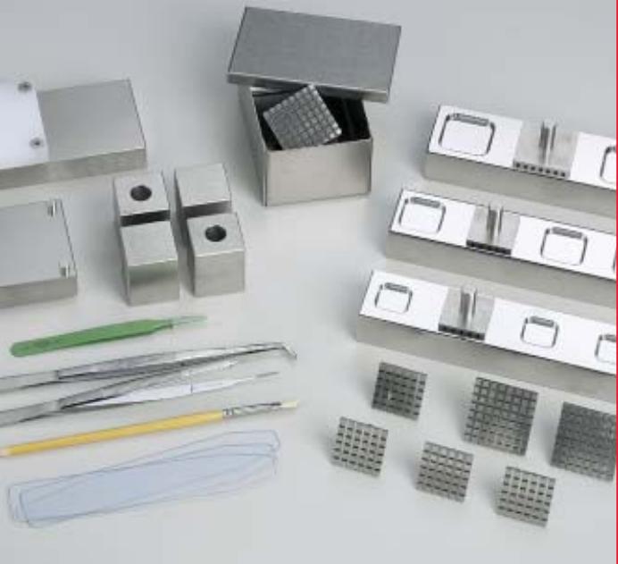
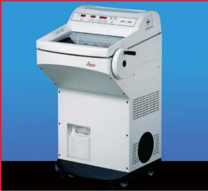
Easily achieve proper specimen orientation and perfect embedding with the unique Precision Cryoembedding System. The Precision System was developed by American pathologist, Dr. Stephen Peters, to expedite and improve frozen sections and shorten the learning process for pathology residents. The system's individual components can be used in a variety of embedding procedures to perfectly embed and properly orient almost any type of specimen. The process is comfortably performed inside the cryostat using stainless steel well bars, chucks, and freezing blocks. Since the Precision Cryoembedding System's components are stored at cold temperatures, most specimens freeze in 20 to 60 seconds, depending upon their size and the selected freezing technique. This significantly reduces turn-around time. And, your well-oriented, flat specimens will be conserved during the trimming process! What else could you ask for?
The Precision Cryoembedding System consists of:
- 3 well bars in 3 sizes (18, 24 and 30 mm )
- 6 small stages (chucks)
4 large stages (chucks)
4 over-stage heat extractors - 1 storage bin for stages
16 dispensing slides - 1 cutting board/freezing griddle
1 elevated heat extractor - 1 pair of angled embedding forceps and accessories
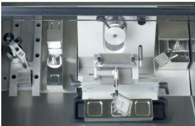
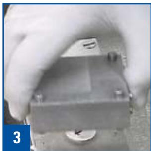
Cryoembedding components in the Leica CM1850 cryochamber
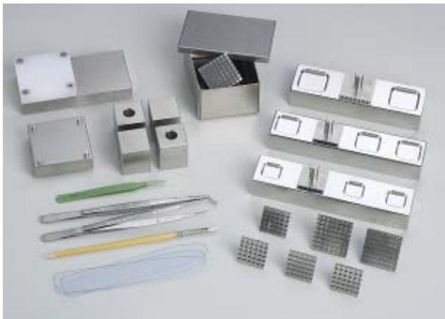
Precision cryoembedding accessories
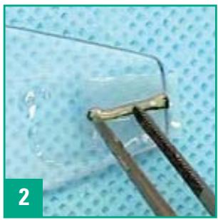
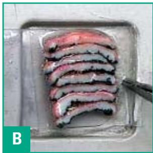
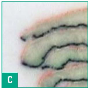
Face Down Cryoembedding
Perfect, Flat Orientation
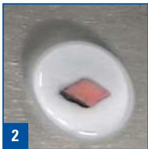
Any specimen, whether it is single or multiple pieces; large or minute; solid or liquid, can be embedded flat and in a single plane.
Technique
- Precisely orient the specimen(s), face down, on a thin plastic dispensing slide.
- Touch the edge of a specimen to the cold base of the embedding well and gently withdraw the dispensing slide while positioning the specimen. Repeat as necessary.
- Slightly overfill the well with embedding medium.
- Place a chuck over the well.
- Place an over-chuck freezing block over the stem of the chuck.
- Remove freezing block and tap the chuck stem when freezing is complete (usually 20 to 60 seconds) to remove the embedded block from the embedding well.
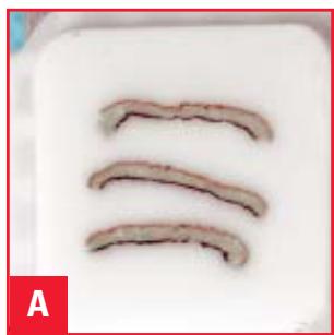
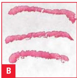
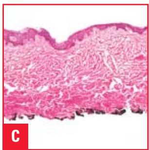
The Results
A: Trimmed block
B: Stained section on slide
C: Photomicrograph
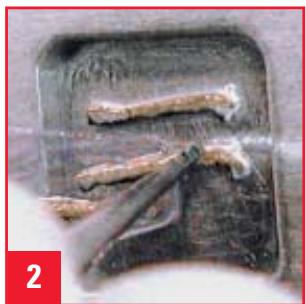
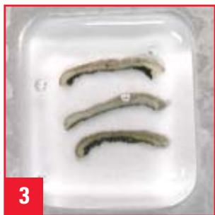
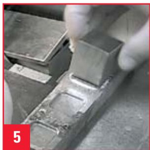
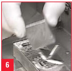
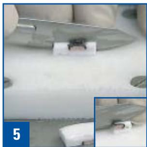
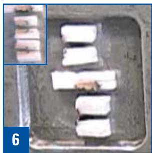
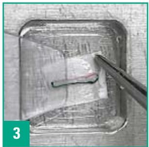
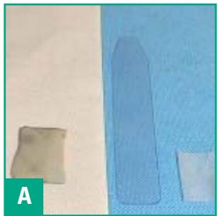
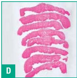
Frozen Block Cryoembedding
Precise, On-edge Orientation
Specimens are embedded and frozen in their entirety, then mapped and cut into firm, flat pieces. The still frozen, flat pieces are then embedded on edge. This technique is perfect for flimsy, tubular, curled, or angular specimens and it is particularly useful for margin resections.
Technique
- Place the specimen face down on the freezing griddle.
- Cover the specimen with a layer of embedding medium.
- Cover the specimen with the appropriate elevated freezing block.
- When completely frozen, map the specimen.
- Cut the embedded block into pieces on the cold cutting board, keeping pieces cold on the adjacent metal surface. (The main photo, #5 to the left, shows the central section while the inset shows a longitudinal margin.)
- Place the cut pieces face down (pieces are face up in the inset photo #6) in the embedding well and freeze using the procedure described under Face Down Cryoembedding.
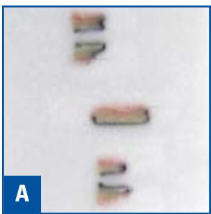
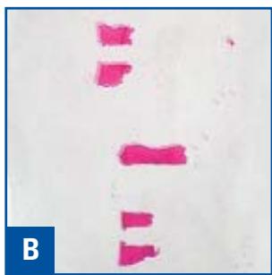
The Results
A: Trimmed block
B: Stained section on slide
C: Photomicrograph (sections repositioned in photograph)
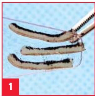
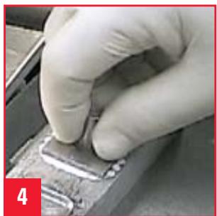
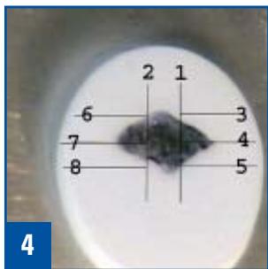
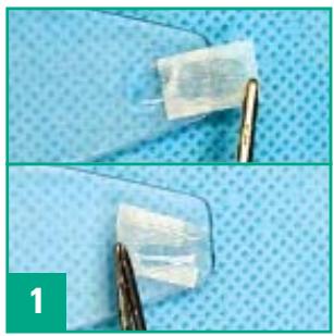
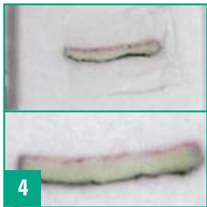
Paper Cryoembedding
Proper Positioning of Difficult Specimens
This technique is used to maintain the orientation of delicate or flimsy specimens or to arrange multiple specimens so they will remain in the same plane for sectioning.
Technique
- Soak a small piece of lens paper in embedding medium and flatten it to the dispensing slide.
- Place the specimen(s) on the lens paper and orient appropriately. Allow an end of the paper to overhang the edge of the dispensing slide.
- Touch the lens paper to the cold floor of the embedding well and gently pull the dispensing slide away. The specimen will remain correctly positioned on the lens paper.
- Trim through the paper on the trimming portion of the blade and then move to a clean, sharp portion to section the specimen. (Untrimmed block in upper portion of photo #4, trimmed block in lower portion.)
The Results
A: Large undissected specimen, dispensing slide and lens paper
B: Specimen dissected with inked border
C: Embedded block
D: Stained sections on slide
Leica Microsystems - the brand for outstanding products
Leica Microsystems' mission is to be the world's first-choice provider of innovative solutions to our customers' needs for vision, measurement, lithography and analysis of microstructures.
Leica, the leading brand for microscopes and scientific instruments, developed from five brand names, all with a long tradition: Wild, Leitz, Reichert, Jung and Cambridge Instruments. Yet Leica symbolizes innovation as well as tradition.
Leica Microsystems – an international company with a strong network of customer services
| Australia: | Gladesville | Tel. +61 2 9879 9700 | Fax +61 2 9817 8358 |
| Austria: | Vienna | Tel. +43 1 486 80 50 0 | Fax +43 1 486 80 50 30 |
| Canada: | Richmond Hill/Ontario | Tel. +1 905 762 2000 | Fax +1 905 762 8937 |
| Denmark: | Herlev | Tel. +45 4454 0101 | Fax +45 4454 0111 |
| France: | Rueil-Malmaison | Tel. +33 1 473 285 85 | Fax +33 1 473 285 86 |
| Germany: | Bensheim | Tel. +49 6251 136 0 | Fax +49 6251 136 155 |
| Italy: | Milan | Tel. +39 0257 486.1 | Fax +39 0257 40 3273 |
| Japan: | Tokyo | Tel. +81 3 5435 9600 | Fax +81 3 5435 9615 |
| Korea: | Seoul | Tel. +82 2 514 65 43 | Fax +82 2 514 65 48 |
| Netherlands: | Rijswijk | Tel. +31 70 4132 100 | Fax +31 70 4132 109 |
| People's Rep. of China: | Hong Kong | Tel. +852 2564 6699 | Fax +852 2564 4163 |
| Portugal: | Lisbon | Tel. +351 21 388 9112 | Fax +351 21 385 4668 |
| Singapore | Tel. +65 6779 7823 | Fax +65 6773 0628 | |
| Spain: | Barcelona | Tel. +34 93 494 95 30 | Fax +34 93 494 95 32 |
| Sweden: | Sollentuna | Tel. +46 8 625 45 45 | Fax +46 8 625 45 10 |
| Switzerland: | Glattbrugg | Tel. +41 1 809 34 34 | Fax +41 1 809 34 44 |
| United Kingdom: | Milton Keynes | Tel. +44 1908 246 246 | Fax +44 1908 609 992 |
| USA: | Bannockburn/llinois | Tel. +1 847 405 0123 | Fax +1 847 405 0164 |
and representatives of Leica Microsystems in more than 100 countries.
The companies of the Leica Microsystems Group operate internationally in four business segments, where we rank with the market leaders.
Microscopy Systems
Our expertise in microscopy is the basis for all our solutions for visualization, measurement and analysis of microstructures in life sciences and industry. With confocal laser technology and image analysis systems, we provide three-dimensional viewing facilities and offer new solutions for cytogenetics, pathology and materials sciences.
- Specimen Preparation
We provide comprehensive systems and services for clinical histo- and cytopathology applications, biomedical research and industrial quality assurance. Our product range includes instruments, systems and consumables for tissue infiltration and embedding, microtomes and cryostats as well as automated stainers and coverslippers.
Medical Equipment
Innovative technologies in our surgical microscopes offer new therapeutic approaches in microsurgery.
- Semiconductor Equipment
Our automated, leading-edge measurement and inspection systems and our E-beam lithography systems make us the first choice supplier for semiconductor manufacturers all over the world.
- Precision
- Cryoembedding System
- Precision Cryoembedding System
- The Precision Cryoembedding System consists of:
- Face Down Cryoembedding
- Perfect, Flat Orientation
- Technique
- The Results
- Frozen Block Cryoembedding
- Precise, On-edge Orientation
- Paper Cryoembedding
- Proper Positioning of Difficult Specimens
- Leica Microsystems - the brand for outstanding products
- Microscopy Systems
- - Specimen Preparation
- Medical Equipment
- - Semiconductor Equipment
Marque : LEICA
Modèle : CRYOEMBEDDING
Catégorie : Équipement de microscopie électronique