
700M - Microscope Levenhuk - Free user manual and instructions
Find the device manual for free 700M Levenhuk in PDF.


User questions about 700M Levenhuk
0 question about this device. Answer the ones you know or ask your own.
Ask a new question about this device
Download the instructions for your Microscope in PDF format for free! Find your manual 700M - Levenhuk and take your electronic device back in hand. On this page are published all the documents necessary for the use of your device. 700M by Levenhuk.
USER MANUAL 700M Levenhuk
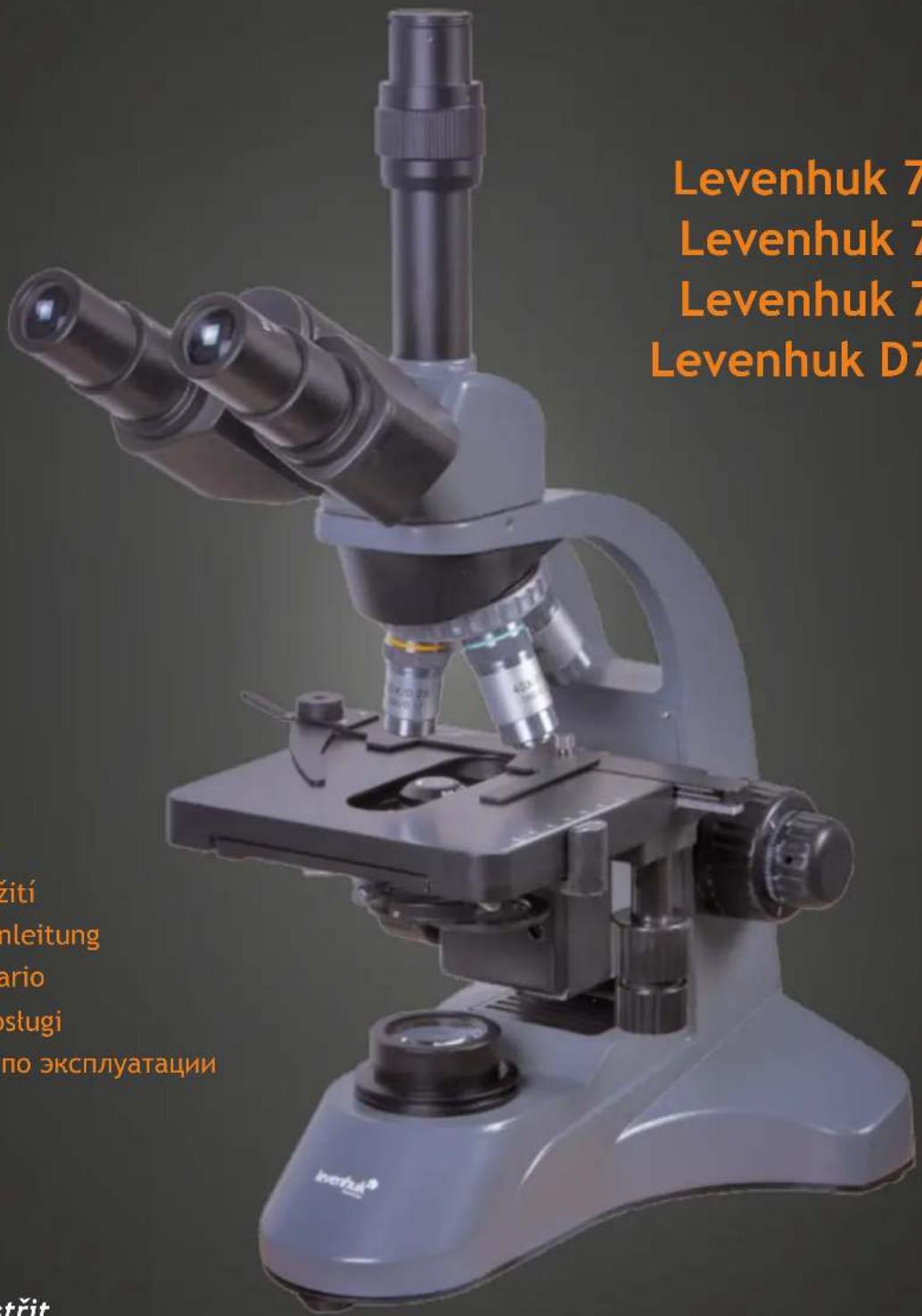
Levenhuk 700 Series Biological Microscopes
Levenhuk 700M
Levenhuk 720B
Levenhuk 740T
Levenhuk D740T
text_image
Levenhuk 7 Levenhuk 7 Levenhuk 7 Levenhuk D7 ziti nleitung ario stugi по эксплуатацииUser Manual
Návod k použití
Bedienungsanleitung
Guía del usuario
Instrukcja obstugi
text_image
Labeled diagram of a microscope with numbered parts for identification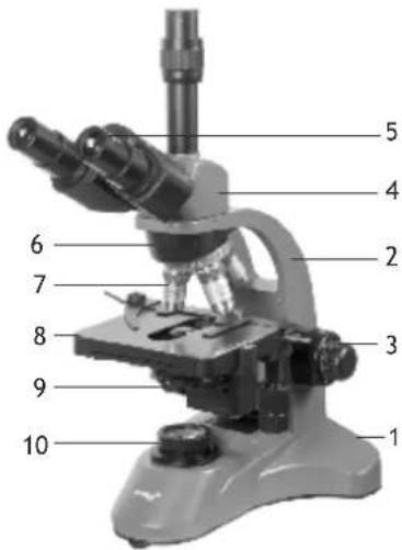
text_image
1 2 3 4 5 6 7 8 9 10EN
1 Base
2 Arm
3 Focus adjustment knob
4 Eyepiece head
5 Eyepieces
6 Revolving nosepiece
7 Objectives
8 Stage
9 Abbe condenser
10 Illumination
11 Brightness adjustment
CZ
Levenhuk 700 Series biological microscopes are modern state-of-the-art optical instruments made with great attention to detail. They are a perfect choice for specialists in all scientific areas. Levenhuk 700 Series microscopes are mainly used for observing and testing biological samples. They perform well during clinical research and tests; teaching demonstrations; in medical and health establishments, laboratories, universities and schools; and may be used for scientific research in agriculture and microbiology.
Package
- microscope body
• eyepiece head: monocular (700M), binocular (720B) or trinocular (740T, D740T) - achromatic objectives: 4x, 10x, 40xs, 100xs (oil)
• eyepieces: WF10x and WFH20x (wide field) - filters: blue, green, yellow
• vial of immersion oil - dust cover
- user manual and warranty
The Levenhuk D740T kit additionally includes: 5Mpx digital camera, a USB cable and a CD with LevenhukLite software & drivers.
Microscope parts
Base. It supports the weight of the microscope, and houses the illumination source, electronics and control mechanisms.
Arm. This piece holds the base, the stage and the head of the microscope together. Coarse and fine focus systems provide for smooth vertical movements of the stage.
Rack-and-pinion mechanism. The stage with the condenser are mounted on the arm and are moving vertically along it. For additional precision, a condenser may be adjusted separately.
Head. A monocular (Levenhuk 700M), binocular (Levenhuk 720B) or a trinocular (Levenhuk 740T and D740T) head is mounted at a 45^ angle at the upper end of the arm and is 360^ rotatable.
Eyepieces and objectives. Consist of lenses that allow magnifying the image. High-quality achromatic objectives with 4x, 10x, 40x and 100x magnifications provide for sharp and bright images. The total magnification is calculated by multiplying the eyepiece magnification to the objective magnification. Thus, the available magnifications are 40x, 100x, 400x and 1000x with the WF10x eyepiece, and 80x, 200x, 800x and 2000x with the WFH20x eyepiece.
Revolving nosepiece. The quadruple revolving nosepiece allows you to change objectives smoothly and easily.
Stage. Sturdy and reliable stage with a specimen holder can be used to move your slides while observing them. The lower illumination light passes through the opening in the middle of the stage.
Condenser. Abbe condenser, 1.25 N.A. iris diaphragm is a system of lenses located below the stage that allows gathering and directing the light rays.
Focusing knob. A coarse and fine focusing system allows moving the stage up and down adjusting the image sharpness.
Lower illumination. LED illumination with adjustable brightness allows observing transparent objects. The illumination system can be powered by A/C.
Operating principle and illumination
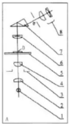
Image creation system: objective (6), prism (7) and eyepiece (9). The objective lens (6) magnifies a specimen (0), light rays pass through the prism (7), refract at a 45^ angle and create an image in the eyepiece. Total magnification may be calculated by multiplying magnifications of the eyepiece and the objective used.
Illumination system: lamp (1), collector lens (2), diaphragm (3) and condenser (4). Light emitted from the lamp (1) passes through the collector lens (2) and illuminates the diaphragm (3). Then, the light is focused by the condenser (4). This illumination system is used for observations of a specimen (0) in transmitted light. You can also use a different type of illumination (e.g. a reflector) for observations in reflected light.
text_image
A 1 2 3 4 5 6 7 8 0Using the microscope
Getting started
- Unpack the microscope and make sure all parts are available.
- Move the stage to the lowermost position using the focusing knob and turn on the illumination or adjust the reflector to evenly illuminate the specimen within the field of view.
- Connect the microscope to A/C power using the A/C adapter.
- Insert the eyepiece into the eyepiece tube.
- Slowly adjust the illumination brightness, from dark to light. When you finish your observations, always dim the illumination before unplugging the power cable.
- If you need to change the light bulb, unplug your microscope first.
Focusing
- Place a specimen on the stage and fix it with the holders.
- Select the 4x objective rotating the revolving nosepiece.
- Center the observed specimen in the field of view.
- Rotate the focusing knob to slowly raise the stage until the objective is close to the specimen; keep checking the distance between the objective and the object to avoid their contact.
CAUTION: The objective should not touch the specimen, otherwise the objective or/and the specimen might be damaged. - Look through the installed eyepiece and lower the stage slowly rotating the focusing knob until you see the sharp image of the observed specimen.
- Such adjustment protects the frontal lens from contacting the object when you use objectives of other magnifications; though, slight refocusing might be required.
- If the image is too bright, adjust the diaphragm disk until the passing light ray is reduced to a comfortable brightness level. If the image is too dark, increase the light ray.
Selecting the objective
- Start your observations with the lowest magnification objective (4x) and select a specimen segment for detailed research. Then move the specimen to center the selected segment in the field of view, to make sure it keeps centered when the objective is changed to a more powerful one. Once the segment is selected, you should center its image in the microscope's field of view as precisely as possible. Otherwise, the desired segment might fail to center in the field of view of the higher power objective.
- Now you can switch to a more powerful objective by rotating the revolving nosepiece. Adjust the image focus, the iris diaphragm and the illumination brightness if required.
- When using a 100x objective lens, you have to fill the space between the lens and the specimen with immersion oil. There should be no air bubbles in the oil, as they might reduce the resulting quality of the image. You must wipe the 100x objective clear after using it.
Digital camera
Levenhuk D740T comes with a 5Mpx digital camera. The digital camera is installed in the eyepiece tube in place of the eyepiece. It camera allows you to observe specimens in fine detail and true colors on your PC monitor and save images on the hard drive. Connect the camera to your PC via a USB cable that also acts as a power cable.
Digital camera specifications:
| Max. resolution (still images) | 2592x1944 |
| Megapixels | 5.1 |
| Sensor | 1/2.5° CMOS |
| Mounting location | eyepiece tube (replaces the eyepiece) |
| Pixel size, μm | 2.2x2.2 |
| Sensitivity, V/lux.sec@550 nm | 0.53 |
| Spectral range | 380–650nm |
| Image format | *.bmp, *.jpg, *.jpeg, *.png, *.tif, *.tiff, *.gif, *.psd, *.ico, *.emf, *.wmf, etc. |
| Video format | *.wmv, *.h264, *.avi, etc. |
| Exposure | ERS (Electronic Rolling Shutter) |
| White balance | auto/manual |
| Exposure control | auto/manual |
| Software features | image size, brightness, exposure control |
| Port | USB 2.0, 480Mb/s |
| System requirements | Windows XP/Vista/7/8/10 (32 and 64 bit), processor up to 2.8GHz Intel Core 2 or higher, USB 2.0 port; also compatible with Mac OS and Linux |
| Software | USB 2.0 driver, LevenhukLite software (image editor) |
| Camera power supply | USB 2.0 cable |
Specifications
| Levenhuk 700M Levenhuk 720B Levenhuk 740T | Levenhuk D740T | ||
| Type | biological | digital, biological | |
| Head | monocular, 360° rota-table, inclined at 45° | binocular, 360° rota-table, inclined at 45° | trinocular, 360° rotatable, inclined at 45° |
| Revolving nosepiece | quadruple | ||
| Magnification | 40x–2000x | ||
| Objectives | achromatic 4x, 10x, 40xs, 100xs (oil) | ||
| Eyepieces | WF10x/18mm, WFH20x | ||
| Optics material | glass | ||
| Body material | aluminum | ||
| Eyepiece tube (length x ∅) | 160mm (6.3in) x 23.2mm (0.9in) | ||
| Stage | double layer mechanical stage, 140x130mm (5.5x5.1in) | ||
| Stage movement range | 24mm vertical, 75mm horizontal | ||
| Condenser | Abbe N.A. 1.25 iris diaphragm and filters | ||
| Focus system | coaxial, coarse (22mm) and fine (0.002mm) | ||
| Illumination | lower (3W LED, brightness adjustment) | ||
| Camera | no | 5Mpx | |
| Power source | AC adapter 220V / 110V 50Hz | ||
Levenhuk reserves the right to modify or discontinue any product without prior notice.
Care and maintenance
- Never, under any circumstances, look directly at the Sun, another bright source of light or at a laser through this device, as this may cause PERMANENT RETINAL DAMAGE and may lead to BLINDNESS.
- Take necessary precautions when using the device with children or others who have not read or who do not fully understand these instructions. After unpacking your microscope and before using it for the first time check for integrity and durability of every component and connection. Do not try to disassemble the device on your own for any reason. For repairs and cleaning of any kind, please contact your local specialized service center.
- Protect the device from sudden impact and excessive mechanical force. Do not apply excessive pressure when adjusting focus. Do not overtighten the locking screws. Do not touch the optical surfaces with your fingers. To clean the device exterior, use only special cleaning wipes and special optics cleaning tools from Levenhuk. Do not use any corrosive or acetone-based fluids to clean the optics. Abrasive particles, such as sand, should not be wiped off lenses, but instead blown off or brushed away with a soft brush. Do not use the device for lengthy periods of time, or leave it unattended in direct sunlight. Keep the device away from water and high humidity. Be careful during your observations, always replace the dust cover after you are finished with observations to protect the device from dust and stains. If you are not using your microscope for extended periods of time, store the objectives and evepieces separately from the microscope.
- Store the device in a dry, cool place away from hazardous acids and other chemicals, away from heaters, open fire and other sources of high temperatures. When using the microscope, try not to use it near flammable materials or substances (benzene, paper, cardboard, plastic, etc.), as the base may heat up during use, and might become a fire hazard. Always unplug the microscope from a power source before opening the base or changing the illumination lamp. Regardless of the lamp type (halogen or incandescent), give it some time to cool down before trying to change it, and always change it to a lamp of the same type. Always use the power supply with the proper voltage, i.e. indicated in the specifications of your new microscope. Plugging the instrument into a different power outlet may damage the electric circuitry of the microscope, burn out the lamp, or even cause a short circuit.
- Seek medical advice immediately if a small part or a battery is swallowed.
Levenhuk International Lifetime Warranty
All Levenhuk telescopes, microscopes, binoculars and other optical products, except for accessories, carry a lifetime warranty against defects in materials and workmanship.
Lifetime warranty is a guarantee on the lifetime of the product on the market.
All Levenhuk accessories are warranted to be free of defects in materials and workmanship for six months from date of retail purchase.
Levenhuk will repair or replace such product or part thereof which, upon inspection by Levenhuk, is found to be defective in materials or workmanship.
As a condition to the obligation of Levenhuk to repair or replace such product, the product must be returned to Levenhuk together with proof of purchase satisfactory to Levenhuk.
This warranty does not cover consumable parts, such as bulbs (electrical, LED, halogen, energy-saving and other types of lamps), batteries (rechargeable and non-rechargeable), electrical consumables etc.
For further details, please visit our web site: https://www.levenhuk.com/warranty
If warranty problems arise, or if you need assistance in using your product, contact the local Levenhuk branch.
Obecné informace
Levenhuk 700M Levenhuk 720B Levenhuk 740T
Levenhuk D740T
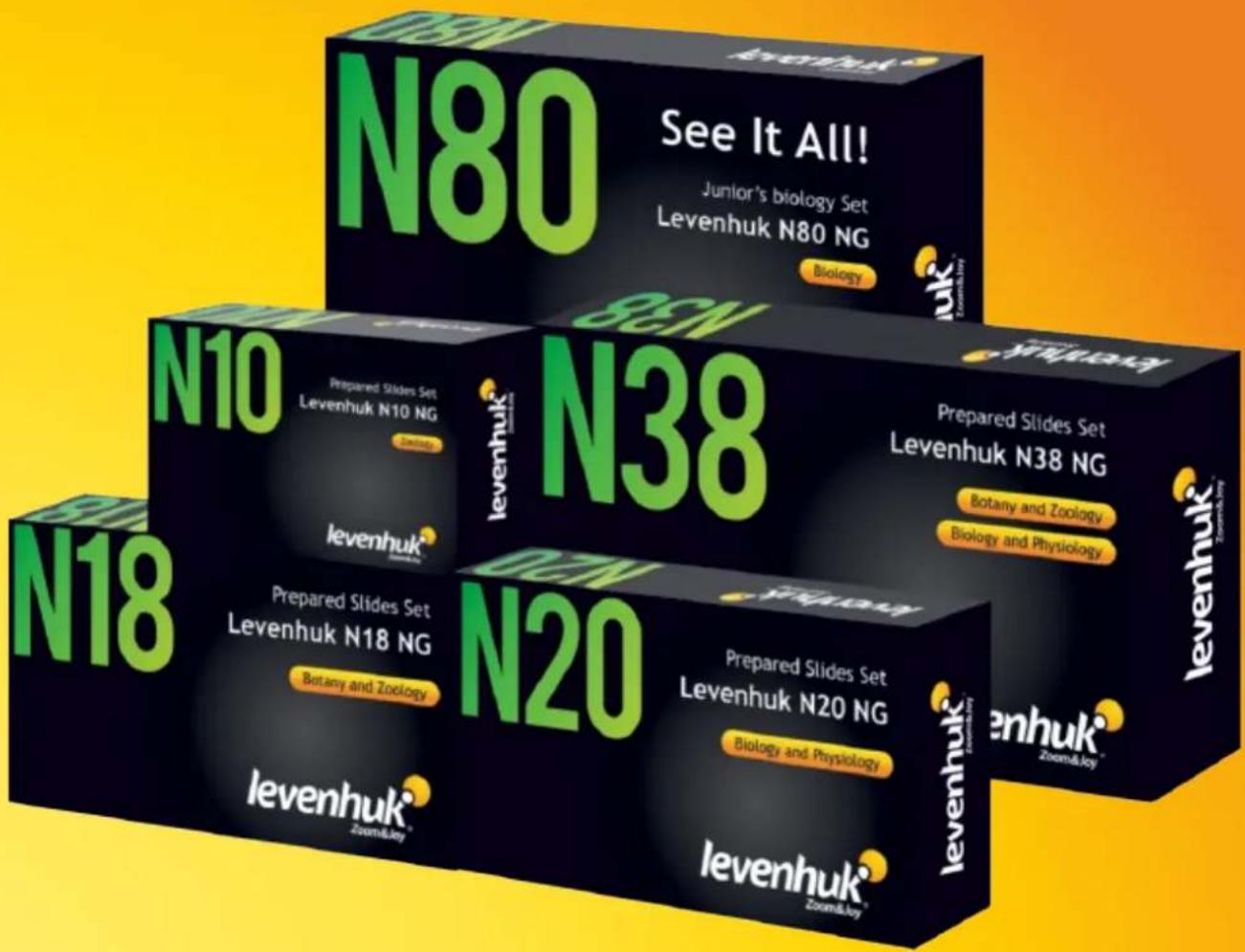
Levenhuk prepared slides sets
The Levenhuk slides sets include thoroughly prepared specimens of various biomaterials as well as blank slides and cover glasses for you to make your own specimens.
Prepared slides, blank slides and cover slips can be used with any microscope model.
text_image
N80 See It All! Junior's biology Set Levenhuk N80 NG Biology N80 N10 Prepared Slides Set Levenhuk N10 NG Zoehnuk® levenhuk® N38 Prepared Slides Set Levenhuk N38 NG Botany and Zoology Biology and Physiology N18 Prepared Slides Set Levenhuk N18 NG Botany and Zoology N20 Prepared Slides Set Levenhuk N20 NG Biology and Physiology levenhuk® Zoom&Joy® levenhuk® Zoom&Joy®Levenhuk Worldwide:
USA: www.levenhuk.com
Bulgaria: www.levenhuk.bg
Canada: ca.levenhuk.com
Czech Rep.: www.levenhuk.cz
Estonia: ee.levenhuk.com
Germany: de.levenhuk.com
Hungary: www.levenhuk.hu
Iceland: is.levenhuk.com Russia: www.levenhuk.ru
India: in.levenhuk.com
Latvia: lv.levenhuk.com
Lithuania: lt.levenhuk.com
Netherlands: nl.levenhuk.com